

 1
172 Best Histology Books of All Time
We've ranked the best histology books using expert recommendations, sales data, and millions of reader ratings. At Shortform, we know books. Our book guides are the best in the world. Learn why.
1 2
2Wheater's Functional Histology: A Text and Colour Atlas
Take a simple approach to understanding the fundamentals with "Wheater's Functional Histology. "Offering concise text accompanied by hundreds of captions and images of histology slides, this best-selling textbook will equip you with all the must-know histology information you need to complete your courses and ace your exams.
 3
3BRS Cell Biology and Histology
Be prepared for your courses and board exams with BRS Cell Biology and Histology ! Acclaimed for its easy-to-read, easy-to-scan outline format, this edition saves time and boosts test-taking confidence. This concise volume distills cell biology and histology facts and concepts commonly covered on course and board exams in a portable, quick-reference format ideal for in-class learning or on-the-go review. Dynamic micrographs, illustrations, full-color tables, and more than 320 board-style multiple-choice review questions with answers reinforce understanding and ensure readiness for exams and the challenges of today’s fast-paced clinical practice.
Updated content reflects the latest clinical perspectives and practices related to cell biology and histology
The proven Board Review Series quick-scan outline format helps students make the most of their study time
Updated high-yield Clinical Considerations boxes demonstrate the practical applications of cell biology and histology concepts
Updated board-style review questions with answers ensure confidence on exams
Full-color tables and high-quality illustrations—including 20 new photomicrographs—clarify concepts and clinical details
Accompanying interactive question bank of questions from the chapter and comprehensive exams ready students for the testing experience
eBook available for purchase. Fast, smart, and convenient, today’s eBooks can transform learning. These interactive, fully searchable tools offer 24/7 access on multiple devices, the ability to highlight and share notes, and more
Updated content reflects the latest clinical perspectives and practices related to cell biology and histology
The proven Board Review Series quick-scan outline format helps students make the most of their study time
Updated high-yield Clinical Considerations boxes demonstrate the practical applications of cell biology and histology concepts
Updated board-style review questions with answers ensure confidence on exams
Full-color tables and high-quality illustrations—including 20 new photomicrographs—clarify concepts and clinical details
Accompanying interactive question bank of questions from the chapter and comprehensive exams ready students for the testing experience
eBook available for purchase. Fast, smart, and convenient, today’s eBooks can transform learning. These interactive, fully searchable tools offer 24/7 access on multiple devices, the ability to highlight and share notes, and more
 4
4Histology: A Text and Atlas: With Correlated Cell and Molecular Biology
Now it its Fifth Edition, this best-selling text and atlas is the perfect text for medical, health professions, and undergraduate biology students. It combines a detailed textbook that emphasizes clinical and functional correlates of histology with a beautifully illustrated atlas featuring full-color digital micrographs of the highest quality.
This edition includes over 100 new illustrations, more Clinical Correlation boxes on the histology of common medical conditions, and new information on the molecular biology of endothelial cell function. Terminology throughout the text is consistent with Terminologia Anatomica.
A powerful interactive histology atlas CD-ROM for students is included with the book and features all of the plates found in the text with interactive functionality.
This edition includes over 100 new illustrations, more Clinical Correlation boxes on the histology of common medical conditions, and new information on the molecular biology of endothelial cell function. Terminology throughout the text is consistent with Terminologia Anatomica.
A powerful interactive histology atlas CD-ROM for students is included with the book and features all of the plates found in the text with interactive functionality.
 5
5Textbook of Histology
Thoroughly updated to reflect all of the latest concepts and advances in the field, this concise, extensively illustrated text presents the basic science and clinical application of cellular and molecular biology. Functioning as a combined text and atlas, it is essentially two books for the price of one providing a comprehensive, visually engaging histology education from the ground up. Illustrations, tables, PowerPointT presentations, chapter summaries and multiple choice questions facilitate comprehension of concepts, and clinical correlations underscore the practical relevance of the material.
Superb illustrations-including photomicrographs, electron micrographs, schematic diagrams and drawings-provide a visual grasp and easier retention of difficult concepts.
Online PowerPointT slides, prepared by the author, offer a convenient way to review the most essential information from the text.
Clinical correlations throughout the text demonstrate clinical applications and reinforce the idea that histology is pertinent not only to pathology and physiology, but in fact comprises one of the essential bases of clinical practice.
New! Pathological Considerations section at the end of each chapter.
Helpful tables provide an at-a-glance summary statement of key points.
Bolded key terms, bulleted lists and chapter summaries emphasize the need to know information in every chapter.
A combination of USMLE-style questions and image based questions in each chapter.
Student Consult eBook version included with purchase. This enhanced eBook experience allows you to search all of the text, figures, and references from the book on a variety of devices.
Superb illustrations-including photomicrographs, electron micrographs, schematic diagrams and drawings-provide a visual grasp and easier retention of difficult concepts.
Online PowerPointT slides, prepared by the author, offer a convenient way to review the most essential information from the text.
Clinical correlations throughout the text demonstrate clinical applications and reinforce the idea that histology is pertinent not only to pathology and physiology, but in fact comprises one of the essential bases of clinical practice.
New! Pathological Considerations section at the end of each chapter.
Helpful tables provide an at-a-glance summary statement of key points.
Bolded key terms, bulleted lists and chapter summaries emphasize the need to know information in every chapter.
A combination of USMLE-style questions and image based questions in each chapter.
Student Consult eBook version included with purchase. This enhanced eBook experience allows you to search all of the text, figures, and references from the book on a variety of devices.
 6
6Pandora's Lab: Seven Stories of Science Gone Wrong
What happens when ideas presented as science lead us in the wrong direction?
History is filled with brilliant ideas that gave rise to disaster, and this book explores the most fascinating—and significant—missteps: from opium's heyday as the pain reliever of choice to recognition of opioids as a major cause of death in the U.S.; from the rise of trans fats as the golden ingredient for tastier, cheaper food to the heart disease epidemic that followed; and from the cries to ban DDT for the sake of the environment to an epidemic-level rise in world malaria.
These are today's sins of science—as deplorable as mistaken past ideas about advocating racial purity or using lobotomies as a cure for mental illness. These unwitting errors add up to seven lessons both cautionary and profound, narrated by renowned author and speaker Paul A. Offit. Offit uses these lessons to investigate how we can separate good science from bad, using some of today's most controversial creations—e-cigarettes, GMOs, drug treatments for ADHD—as case studies. For every "Aha!" moment that should have been an "Oh no," this book is an engrossing account of how science has been misused disastrously—and how we can learn to use its power for good.
History is filled with brilliant ideas that gave rise to disaster, and this book explores the most fascinating—and significant—missteps: from opium's heyday as the pain reliever of choice to recognition of opioids as a major cause of death in the U.S.; from the rise of trans fats as the golden ingredient for tastier, cheaper food to the heart disease epidemic that followed; and from the cries to ban DDT for the sake of the environment to an epidemic-level rise in world malaria.
These are today's sins of science—as deplorable as mistaken past ideas about advocating racial purity or using lobotomies as a cure for mental illness. These unwitting errors add up to seven lessons both cautionary and profound, narrated by renowned author and speaker Paul A. Offit. Offit uses these lessons to investigate how we can separate good science from bad, using some of today's most controversial creations—e-cigarettes, GMOs, drug treatments for ADHD—as case studies. For every "Aha!" moment that should have been an "Oh no," this book is an engrossing account of how science has been misused disastrously—and how we can learn to use its power for good.
 7
7Color Atlas and Text of Histology
Magnify your understanding of histology with Color Atlas and Text of Histology --the student’s go-to tool for histology mastery!
The Seventh Edition of this text and atlas combines traditional topics with modern research to provide medical, dental, allied health, and biology students with a concise review of all of the major tissue classes and body systems. Consistent presentation of histologic plates comprising relevant concise text and photomicrographs facilitates understanding as well as review both for in-class and licensing examinations. Helpful explanatory text in each chapter includes Introductions, new Histophysiology sections, Clinical Considerations, and Summaries of Histological Organization. Numerous tables have also been added to make the material easier to grasp. This atlas features more than 700 vivid, full-color and high-quality photomicrographs, scanning electron micrographs, and schematic illustrations to give students the full histologic picture.
New to the seventh edition:
Revised and updated text ensures that each chapter offers the most current histology and related concepts.
Reorganized chapter content clearly delineates morphology and histophysiology sections.
Expanded Histologic Techniques chapter elucidates various stains and techniques.
Abundant tables organize related topics for quick, easy absorption and review.
Clinical Considerations with clinical images demonstrate the link between histology and pathophysiology.
Full-page Graphics provide a representational orientation before zooming in on micrographs.
Helpful full-color thumbnail drawings complement micrographs for enhanced interpretation.
New appendix helps students distinguish between tissues that resemble each other.
New Review Plates at the end of each chapter.
An interactive question bank of multiple-choice review questions is available for students via thePoint.
The Seventh Edition of this text and atlas combines traditional topics with modern research to provide medical, dental, allied health, and biology students with a concise review of all of the major tissue classes and body systems. Consistent presentation of histologic plates comprising relevant concise text and photomicrographs facilitates understanding as well as review both for in-class and licensing examinations. Helpful explanatory text in each chapter includes Introductions, new Histophysiology sections, Clinical Considerations, and Summaries of Histological Organization. Numerous tables have also been added to make the material easier to grasp. This atlas features more than 700 vivid, full-color and high-quality photomicrographs, scanning electron micrographs, and schematic illustrations to give students the full histologic picture.
New to the seventh edition:
Revised and updated text ensures that each chapter offers the most current histology and related concepts.
Reorganized chapter content clearly delineates morphology and histophysiology sections.
Expanded Histologic Techniques chapter elucidates various stains and techniques.
Abundant tables organize related topics for quick, easy absorption and review.
Clinical Considerations with clinical images demonstrate the link between histology and pathophysiology.
Full-page Graphics provide a representational orientation before zooming in on micrographs.
Helpful full-color thumbnail drawings complement micrographs for enhanced interpretation.
New appendix helps students distinguish between tissues that resemble each other.
New Review Plates at the end of each chapter.
An interactive question bank of multiple-choice review questions is available for students via thePoint.
 8
8Junqueira's Basic Histology: Text and Atlas, Fifteenth Edition
Publisher's Note: Products purchased from Third Party sellers are not guaranteed by the publisher for quality, authenticity, or access to any online entitlements included with the product.
The text that has defined histology for generations--concise, clear, beautifully illustrated, and better than ever
A Doody's Core Title for 2019!
For more than four decades, Junqueira's Basic Histology has built a global reputation as the most accessible, yet comprehensive overview of human tissue structure and function available. This trusted classic delivers a well-organized and concise presentation of cell biology and histology that integrates the material with that of biochemistry, immunology, endocrinology, and physiology, and provides an excellent foundation for subsequent studies in pathology. Junqueira's is written specifically for students of medicine and other health-related professions, as well as for advanced undergraduate courses in tissue biology - and there is nothing else like it.
FEATURES
-Electron and light micrographs comprise a definitive atlas of cell, tissue, and organ structures
-NEW! Each chapter now includes a set of multiple-choice self-test questions that allow you to assess your comprehension of important material, with some questions utilizing clinical vignettes or cases to provide real-world relevance
-Summary of Key Points and Summary Tables highlight what is important and present it in a way that makes it memorable
-Streamlined page design --including concise, high-yield paragraphs; bullets; and bolded key terms
-Acclaimed art and other figures facilitate learning and visualization of key aspects of cell biology and histology
-A cohesive organization examines how to study the structures of cells and tissues; the cell cytoplasm and nucleus; and the four basic tissue types and their role in the organ systems
-Clinical Correlations presented with each topic
-All-inclusive coverage encompasses all tissues, every organ system, organs, bone and cartilage, blood, skin, and more
-Valuable appendix on light microscopy stains clearly explains this need-to-know staining technique
The text that has defined histology for generations--concise, clear, beautifully illustrated, and better than ever
A Doody's Core Title for 2019!
For more than four decades, Junqueira's Basic Histology has built a global reputation as the most accessible, yet comprehensive overview of human tissue structure and function available. This trusted classic delivers a well-organized and concise presentation of cell biology and histology that integrates the material with that of biochemistry, immunology, endocrinology, and physiology, and provides an excellent foundation for subsequent studies in pathology. Junqueira's is written specifically for students of medicine and other health-related professions, as well as for advanced undergraduate courses in tissue biology - and there is nothing else like it.
FEATURES
-Electron and light micrographs comprise a definitive atlas of cell, tissue, and organ structures
-NEW! Each chapter now includes a set of multiple-choice self-test questions that allow you to assess your comprehension of important material, with some questions utilizing clinical vignettes or cases to provide real-world relevance
-Summary of Key Points and Summary Tables highlight what is important and present it in a way that makes it memorable
-Streamlined page design --including concise, high-yield paragraphs; bullets; and bolded key terms
-Acclaimed art and other figures facilitate learning and visualization of key aspects of cell biology and histology
-A cohesive organization examines how to study the structures of cells and tissues; the cell cytoplasm and nucleus; and the four basic tissue types and their role in the organ systems
-Clinical Correlations presented with each topic
-All-inclusive coverage encompasses all tissues, every organ system, organs, bone and cartilage, blood, skin, and more
-Valuable appendix on light microscopy stains clearly explains this need-to-know staining technique
 9
9Junqueira's Basic Histology: Text and Atlas
The most authoritative, current, and beautifully illustrated histology text available--NEW chapter-ending multiple-choice questions review must-know material
A Doody's Core Title for 2017!
NEW clinical vignettes have been added to each chapter
Full-color, easy-to-understand drawings provide just the right level of detail necessary to reinforce key concepts and facilitate comprehension and retention of text material
For more than three decades, Junqueira's Basic Histology has been unmatched in its ability to explain the function of cell and tissue structure in the human body. Updated to reflect the latest research in the field, and enhanced with more than 1,000 illustrations, most in full-color, the Fourteenth Edition reflects the most comprehensive and modern approach to understanding medical histology available anywhere. This well-regarded classic is distinguished by chapters focusing on the cytoplasmic and nuclear compartment of the cell, the four basic tissues that form the organs, and each organ system.
In response to reader demand, the legends are now concise, stand-alone summaries of the illustrations. Applauded for its visual appeal, Junqueira's is enhanced by full-color micrographs that comprise a complete atlas of tissue sections. These state-of-the-art micrographs highlight the important features of every tissue and organ in the human body, while full-color, easy-to-understand drawings provide just the right level of detail necessary to clarify the text and make learning easier.
A Doody's Core Title for 2017!
NEW clinical vignettes have been added to each chapter
Full-color, easy-to-understand drawings provide just the right level of detail necessary to reinforce key concepts and facilitate comprehension and retention of text material
For more than three decades, Junqueira's Basic Histology has been unmatched in its ability to explain the function of cell and tissue structure in the human body. Updated to reflect the latest research in the field, and enhanced with more than 1,000 illustrations, most in full-color, the Fourteenth Edition reflects the most comprehensive and modern approach to understanding medical histology available anywhere. This well-regarded classic is distinguished by chapters focusing on the cytoplasmic and nuclear compartment of the cell, the four basic tissues that form the organs, and each organ system.
In response to reader demand, the legends are now concise, stand-alone summaries of the illustrations. Applauded for its visual appeal, Junqueira's is enhanced by full-color micrographs that comprise a complete atlas of tissue sections. These state-of-the-art micrographs highlight the important features of every tissue and organ in the human body, while full-color, easy-to-understand drawings provide just the right level of detail necessary to clarify the text and make learning easier.
 10
10Histology: A Text and Atlas: With Correlated Cell and Molecular Biology
Now in its seventh edition, Histology: A Text and Atlas is ideal for medical, dental, health professions, and undergraduate biology and cell biology students. This best-selling combination text and atlas includes a detailed textbook, which emphasizes clinical and functional correlates of histology fully supplemented by vividly informative illustrations and photomicrographs. Separate, superbly illustrated atlas sections follow almost every chapter and feature large-size, full-color digital photomicrographs with labels and accompanied descriptions that highlight structural and functional details of cells, tissues, and organs.
Updated throughout to reflect the latest advances in the field, this “two in one” text and atlas features an outstanding art program with all illustrations completely revised and redrawn as well as a reader-friendly format including red highlighted key terms, blue clinical text, and folders that cover clinical correlations and functional considerations.
NEW! All illustrations are now completely revised and redrawn for a consistent art program.
NEW! Histology 101 sections provide students with a reader-friendly review of essential information covered in the preceding chapters.
NEW! Updated cellular and molecular biology coverage reflects the latest advances in the field.
More than 100 atlas plates that incorporate 435 full-color, high-resolution photomicrographs.
Reader-friendly highlights including red bold terms, blue clinical text, and folders featuring clinical and functional correlations that increase student understanding and facilitates efficient study.
Easy-to-understand tables aid students in learning and reviewing information (such as staining techniques) without having to rely on rote memorization.
Features of cells, tissues, and organs and their functions and locations are presented in easy-to-locate, easy-to-review bulleted lists.
Additional clinical correlation and functional consideration folders have been added providing information related to symptoms, photomicrographs of diseased tissues or organs, short histopathological descriptions, and molecular basis for clinical intervention.
Updated throughout to reflect the latest advances in the field, this “two in one” text and atlas features an outstanding art program with all illustrations completely revised and redrawn as well as a reader-friendly format including red highlighted key terms, blue clinical text, and folders that cover clinical correlations and functional considerations.
NEW! All illustrations are now completely revised and redrawn for a consistent art program.
NEW! Histology 101 sections provide students with a reader-friendly review of essential information covered in the preceding chapters.
NEW! Updated cellular and molecular biology coverage reflects the latest advances in the field.
More than 100 atlas plates that incorporate 435 full-color, high-resolution photomicrographs.
Reader-friendly highlights including red bold terms, blue clinical text, and folders featuring clinical and functional correlations that increase student understanding and facilitates efficient study.
Easy-to-understand tables aid students in learning and reviewing information (such as staining techniques) without having to rely on rote memorization.
Features of cells, tissues, and organs and their functions and locations are presented in easy-to-locate, easy-to-review bulleted lists.
Additional clinical correlation and functional consideration folders have been added providing information related to symptoms, photomicrographs of diseased tissues or organs, short histopathological descriptions, and molecular basis for clinical intervention.
 11
11Histology: A Text and Atlas: With Correlated Cell and Molecular Biology
 12
12Junqueira's Basic Histology: Text and Atlas, Thirteenth Edition
The histology text the medical field turns to first -- authoritative, concise, beautifully illustrated, and completely up-to-date
"More than 600 full-color illustrations"
For more than three decades, "Junquiera's Basic Histology" has been unmatched in its ability to explain the relationship between cell and tissue structure with their function in the human body. Updated to reflect the latest research in the field and enhanced with more than 600 full-color illustrations, the thirteenh edition of "Junqueira's" represents the most comprehensive and modern approach to understanding medical histology available anywhere.
"More than 600 full-color illustrations"
For more than three decades, "Junquiera's Basic Histology" has been unmatched in its ability to explain the relationship between cell and tissue structure with their function in the human body. Updated to reflect the latest research in the field and enhanced with more than 600 full-color illustrations, the thirteenh edition of "Junqueira's" represents the most comprehensive and modern approach to understanding medical histology available anywhere.
 13
13Difiore's Atlas of Histology with Functional Correlations
diFiore's Atlas of Histology with Functional Correlations, Eleventh Edition, explains basic histology concepts through full-color, schematic illustrations. These illustrations are supplemented by more than 450 digitized full-color online photomicrographs of histological images. Part One explains tissues and their relationship to their systems; Part Two addresses organs in a similar way.
Targeting undergraduate, allied health and first and second year medical students, the Eleventh Edition includes new and enhanced images through redrawing and digitization to provide increased detail. This edition also features updated illustrations and information on the functions of cells, tissues, and organs of the body based on advances in research and expert recommendations.
The atlas' student-friendly "Functional Correlations" sections help students study structure and function together. Students also benefit from a "realistic" perspective as more than 70 micrographs appear adjacent to color illustrations.
A companion Website offers student and instructor versions of diFiore's Interactive Atlas with all of the images from the book.
Targeting undergraduate, allied health and first and second year medical students, the Eleventh Edition includes new and enhanced images through redrawing and digitization to provide increased detail. This edition also features updated illustrations and information on the functions of cells, tissues, and organs of the body based on advances in research and expert recommendations.
The atlas' student-friendly "Functional Correlations" sections help students study structure and function together. Students also benefit from a "realistic" perspective as more than 70 micrographs appear adjacent to color illustrations.
A companion Website offers student and instructor versions of diFiore's Interactive Atlas with all of the images from the book.
 14
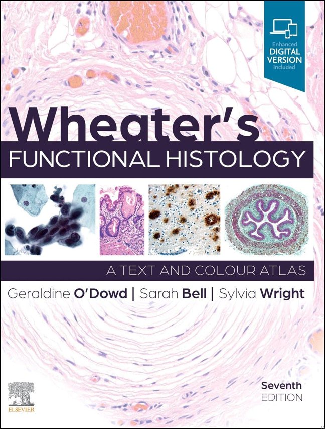
14Wheater's Functional Histology
**Selected for 2026 Doody's Core Titles in Cell Biology/Histology**Now in its seventh edition, Wheater's Functional Histology is designed to give students a working knowledge of histology, including the basic histological structures, tissue types and organ systems.You will learn to recognise the microscopic structure of normal human tissues and understand how this relates to function. The book shows you how to apply histology in a clinical context through coverage of common clinical conditions in each chapter.This must-have book is written clearly and concisely, designed for those studying histology for the first time. Packed with a wealth of illustrations and learning aids, it is a must-have for every medical and health science student.Concise and easy to digest text – easy to follow for those new to histologyMore than 900 high quality histology images and illustrations, plus detailed explanations to support understandingEnd of chapter review tables summarising important pointsSelf-assessment questions and rationales to support exam preparationAppendix outlining the basics of microscopy and histological staining techniquesBasic glossaryAn enhanced eBook version is included with purchase. The eBook allows you to access all the text, figures and references, with the ability to search, customize your content, make notes and highlights, and have content read aloud
 15
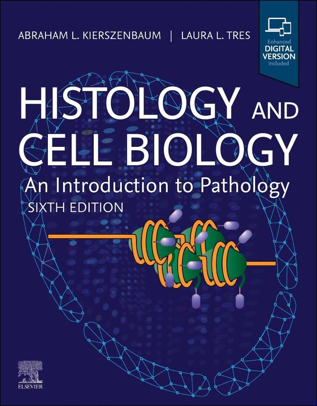
15Histology and Cell Biology: An Introduction to Pathology
The first text to integrate microscopic anatomy with cell biology, general pathology, and physiology, Histology and Cell Biology: An Introduction to Pathology, Sixth Edition, continues to set the standard for studying today’s rapidly evolving, molecular-oriented medicine. Drs. Abraham L. Kierszenbaum and Laura L. Tres present essential concepts in an accessible, easy-to-understand manner, with full-color illustrations, diagrams, photomicrographs, and pathology photos fully integrated on every page. Linking pathological conditions to basic sciences throughout, this award-winning text helps you build a substantial clinical knowledge base while learning the structure and function of normal organs.Provides cover-to-cover updates throughout, reflecting current terminology, basic science, and clinical conceptsIncludes new material at the beginning of each chapter that assists you with the microscope as you first encounter the architectural complexity of tissues or organsIncludes Primers in most chapters that provide a practical, self-contained integration of histology, cell biology, and pathology, helping you understand the relationship between basic and clinical sciencesContains Essential Concepts at the end of each chapter highlighting relevant issues―perfect for studying and reviewing for in-course and board examinationsUses a light green background throughout the text to identify essential concepts of histology―a feature requested by both students and instructors to quickly locate which concepts are most important for beginning learners or when time is limitedIdentifies clinical terms throughout the text and lists all clinical boxes in the table of contents for quick referenceHelps you understand the links between chapter concepts with concept mapping animations online, providing a dynamic supplement to in-class instructionAn eBook version is included with purchase. The eBook allows you to access all of the text and figures, with the ability to search, customize your content, make notes and highlights, and have content read aloud
 16
16Internal Medicine: Essential Clinical Foundations: Fifth Edition: Evidence-Based Diagnosis, Clinical Management, Case-Based Learning, and Board Review for Medical Trainees
Build the Clinical Foundation You Need Before the Pressure BeginsInternal medicine requires more than memorizing facts. It requires the ability to connect symptoms, diagnostic reasoning, evidence-based treatment, pharmacology, pathology, and patient care into one clear clinical approach.For many medical trainees, the challenge is not a lack of information. It is having too much information scattered across dense textbooks, lecture notes, drug references, and review materials that do not always translate into practical decision-making.This book was created to make that foundation clearer, stronger, and easier to apply.A Practical, High-Yield Guide for Medical Trainees and Clinical ReviewInternal Medicine: Essential Clinical Foundations is a structured academic reference designed to help students and trainees understand the core principles of adult medicine with clarity, confidence, and clinical relevance.It brings together essential concepts across diagnosis, management, pharmacology, case-based learning, and board-style review, including:Evidence-based diagnostic reasoning and clinical assessmentCommon adult medical conditions across major organ systemsMedication principles, therapeutic decision-making, and safety considerationsPathophysiology and histologic foundations linked to clinical diseaseCase-based learning for applied medical reasoningBoard-style review concepts for exam preparationPractical management frameworks for real clinical scenariosEach topic is organized to support both deep learning and efficient review.Built for Speed, Designed for UseThis fifth edition is structured for readers who need a dependable resource that is clear, organized, and practical.System-based organization for faster navigationConcise explanations of essential mechanisms and clinical findingsHigh-yield diagnostic and management frameworksCase-based examples that strengthen applied reasoningMedication-focused guidance for safer clinical decision-makingTables, summaries, and review points for efficient studyBoard-oriented reinforcement without unnecessary overloadThe goal is simple: help you understand what matters, remember it more effectively, and use it with greater confidence.Who This Book Is ForThis resource is designed for learners and professionals building or reviewing a strong internal medicine foundation:Medical students preparing for clinical rotations and examsResidents and early-career clinicians strengthening diagnostic reasoningNursing students and nurses reviewing pharmacology and adult medical careAllied health students needing a structured medical referenceBoard review candidates seeking clear, high-yield reinforcementWhy It WorksThis is not a scattered review book or an overwhelming specialty textbook.It is built around how internal medicine is actually learned and applied:Clinically organized, so concepts connect to patient careEvidence-based, so decisions are grounded in modern standardsReview-friendly, so complex material becomes easier to retainIt helps bridge the gap between academic knowledge and practical clinical judgment.Stay Ready—Even Under PressureWhether you are preparing for exams, reviewing before rounds, strengthening pharmacology knowledge, or building confidence in patient assessment, this guide gives you a clear framework for learning internal medicine with purpose.Structured. Practical. Built for modern medical training.Add it to your collection and build the clinical foundation you can rely on when it matters most.
 17
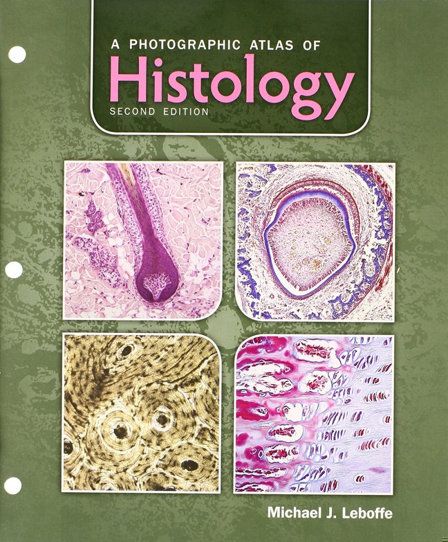
17A Photographic Atlas of Histology
Includes bibliographical references and index.
Cover unavailable
18Color Textbook of Histology
The new edition of this well-received text provides students with concise, current coverage of cell and molecular biology and classic morphology. It features a wealth of superb illustrations, including full-color light photomicrographs, electron micrographs, schematic diagrams, and drawings. Helpful tables summarize vital information for at-a-glance understanding, and summary statements highlight key material in each chapter.
Cover unavailable
19Color Textbook of Histology 3RD EDITION
 20
20Lippincott's Illustrated Q Review of Histology
Lippincott’s Illustrated Q&A Review of Histology is a resource for students engaged in histology course review and test preparation for the USMLE Step 1 and COMLEX. It contains more than 1,000 USMLE-style and content recall questions with images for approximately 40% of the questions.
Features:
USMLE-style multiple choice questions—each vignette is followed by a question stem that addresses a key concept in cell biology/histology
Detailed explanations address incorrect answers in context
Over 480 full-color images illustrate important histologic features
Tissues with similar histological features are compared, providing a challenging comprehensive review
Side-by-side comparisons of normal tissue and histopathology provide a bridge to clinical problem solving and diagnostic pathology
Online access to the questions and answers provides flexible study options
Features:
USMLE-style multiple choice questions—each vignette is followed by a question stem that addresses a key concept in cell biology/histology
Detailed explanations address incorrect answers in context
Over 480 full-color images illustrate important histologic features
Tissues with similar histological features are compared, providing a challenging comprehensive review
Side-by-side comparisons of normal tissue and histopathology provide a bridge to clinical problem solving and diagnostic pathology
Online access to the questions and answers provides flexible study options
Cover unavailable
21The Poison Squad: One Chemist's Single-Minded Crusade for Food Safety at the Turn of the Twentieth Century
A New York Times Notable Book
From Pulitzer Prize winner and New York Times-bestselling author Deborah Blum, the dramatic true story of how food was made safe in the United States and the heroes, led by the inimitable Dr. Harvey Washington Wiley, who fought for change
By the end of nineteenth century, food was dangerous. Lethal, even. "Milk" might contain formaldehyde, most often used to embalm corpses. Decaying meat was preserved with both salicylic acid, a pharmaceutical chemical, and borax, a compound first identified as a cleaning product. This was not by accident; food manufacturers had rushed to embrace the rise of industrial chemistry, and were knowingly selling harmful products. Unchecked by government regulation, basic safety, or even labelling requirements, they put profit before the health of their customers. By some estimates, in New York City alone, thousands of children were killed by "embalmed milk" every year. Citizens--activists, journalists, scientists, and women's groups--began agitating for change. But even as protective measures were enacted in Europe, American corporations blocked even modest regulations. Then, in 1883, Dr. Harvey Washington Wiley, a chemistry professor from Purdue University, was named chief chemist of the agriculture department, and the agency began methodically investigating food and drink fraud, even conducting shocking human tests on groups of young men who came to be known as, "The Poison Squad."
Over the next thirty years, a titanic struggle took place, with the courageous and fascinating Dr. Wiley campaigning indefatigably for food safety and consumer protection. Together with a gallant cast, including the muckraking reporter Upton Sinclair, whose fiction revealed the horrific truth about the Chicago stockyards; Fannie Farmer, then the most famous cookbook author in the country; and Henry J. Heinz, one of the few food producers who actively advocated for pure food, Dr. Wiley changed history. When the landmark 1906 Food and Drug Act was finally passed, it was known across the land, as "Dr. Wiley's Law."
Blum brings to life this timeless and hugely satisfying "David and Goliath" tale with righteous verve and style, driving home the moral imperative of confronting corporate greed and government corruption with a bracing clarity, which speaks resoundingly to the enormous social and political challenges we face today.
From Pulitzer Prize winner and New York Times-bestselling author Deborah Blum, the dramatic true story of how food was made safe in the United States and the heroes, led by the inimitable Dr. Harvey Washington Wiley, who fought for change
By the end of nineteenth century, food was dangerous. Lethal, even. "Milk" might contain formaldehyde, most often used to embalm corpses. Decaying meat was preserved with both salicylic acid, a pharmaceutical chemical, and borax, a compound first identified as a cleaning product. This was not by accident; food manufacturers had rushed to embrace the rise of industrial chemistry, and were knowingly selling harmful products. Unchecked by government regulation, basic safety, or even labelling requirements, they put profit before the health of their customers. By some estimates, in New York City alone, thousands of children were killed by "embalmed milk" every year. Citizens--activists, journalists, scientists, and women's groups--began agitating for change. But even as protective measures were enacted in Europe, American corporations blocked even modest regulations. Then, in 1883, Dr. Harvey Washington Wiley, a chemistry professor from Purdue University, was named chief chemist of the agriculture department, and the agency began methodically investigating food and drink fraud, even conducting shocking human tests on groups of young men who came to be known as, "The Poison Squad."
Over the next thirty years, a titanic struggle took place, with the courageous and fascinating Dr. Wiley campaigning indefatigably for food safety and consumer protection. Together with a gallant cast, including the muckraking reporter Upton Sinclair, whose fiction revealed the horrific truth about the Chicago stockyards; Fannie Farmer, then the most famous cookbook author in the country; and Henry J. Heinz, one of the few food producers who actively advocated for pure food, Dr. Wiley changed history. When the landmark 1906 Food and Drug Act was finally passed, it was known across the land, as "Dr. Wiley's Law."
Blum brings to life this timeless and hugely satisfying "David and Goliath" tale with righteous verve and style, driving home the moral imperative of confronting corporate greed and government corruption with a bracing clarity, which speaks resoundingly to the enormous social and political challenges we face today.
 22
22Lippincott Illustrated Reviews: Cell and Molecular Biology (Lippincott Illustrated Reviews Series)
Up to date, easy to use, and rich with vibrant illustrations, Lippincott® Illustrated Reviews: Cell and Molecular Biology, 3rd Edition, provides a highly visual presentation of essential cell and molecular biology with a focus on topics related to human health and disease. This engaging approach incorporates all of the most popular features of the bestselling Lippincott® Illustrated Reviews series, including abundant full-color illustrations, chapter summaries, and review questions that link basic science to real-life clinical situations. The updated, versatile 3rd Edition can be used for a standalone cell biology course in medical, health professions, or other graduate and upper-level undergraduate programs; as a review for course and board exams; or in conjunction with other Lippincott® Illustrated Reviews for a seamless integrated course.
Cover unavailable
23An Atlas of Histology
The beginning student of histology is frequently confronted by a paradox: diagrams in many books that illustrate human microanatomy in a simplified, cartoon-like manner are easy to understand, but are difficult to relate to actual tissue specimens or photographs. In turn, photographs often fail to show some important features of a given tissue, because no individual specimen can show all of the tissue's salient fea- tures equally well. This atlas, filled with photo-realistic drawings, was prepared to help bridge the gap between the simplicity of diagrams and the more complex real- ity of microstructure. All of the figures in this atlas were drawn from histological preparations used by students in my histology classes, at the level of light microscopy. Each drawing is not simply a depiction of an individual histological section, but is also a synthesis of the key structures and features seen in many preparations of similar tissues or organs. The illustrations are representative of the typical features of each tissue and organ. The atlas serves as a compendium of the basic morphological characteristics of human tissue which students should be able to recognize.
 24
24Cajal's Butterflies of the Soul: Science and Art
This book contains a large collection of beautiful figures produced throughout the nineteenth century and the beginning of the twentieth century and that represent some characteristic examples of the early days of research in neuroscience. The main aim of this work is to demonstrate to the general public that the study of the nervous system is not only important for the many obvious reasons related to brain function in both health and disease, but also for the unexpected natural beauty that it beholds. This beauty has been discovered thanks to the techniques used to visualize the microscopic structure of the brain, a true forest of colorful and florid neural cells. As illustrated by his marvelous drawings, the studies of Santiago Ram�n y Cajal (1852-1934) no doubt contributed more than those of any other researcher at the time to the growth of modern neuroscience. Thus, we have honored his name in the title of this book, even though the figures contained in the main body of the book are from 91 authors. Looking at the illustrations in this book the readers will not only marvel at Cajal's drawings but they will also find that many of the other early researchers that studied the nervous system were also true artists, of considerable talent and aesthetic sensibility. Thus, the present book contains numerous drawings of some of the most important pioneers in neuroscience, including Deiters, K�lliker, Meynert, Ranvier, Golgi, Retzius, Nissl, Dogiel, Alzheimer, del R�o-Hortega and de Castro.
The book has been divided into two Parts, Part I and II, the latter containing the main body of the work. Part I contains introductory information that will give readers unfamiliar with the nervous system a better understanding of the importance of the scientific illustrations produced in those days. The second part of the book, Part II, contains the collection of 282 figures with the intention of transforming the reader into an observer. These illustrations have been divided in three main categories: Section I, The Benedictine period: The early days; Section II, The black period: Neurons, glia and organization of the nervous system; Section III, The colorful period: internal structure and chemistry of the cells.
This book will be of general interest, not only due to the captivating aesthetic appeal of the illustrations but also because they represent the bases of our current understanding of the nervous system. The reader will find that many of the illustrations can be considered to belong to different artistic movements, such as modernism, surrealism, cubism, abstract art or impressionism. Indeed, these illustrations may also provide artists with a source of inspiration since they reveal a fantastic and virtually unknown world of forms, a microuniverse with an aura of mystery.
The book has been divided into two Parts, Part I and II, the latter containing the main body of the work. Part I contains introductory information that will give readers unfamiliar with the nervous system a better understanding of the importance of the scientific illustrations produced in those days. The second part of the book, Part II, contains the collection of 282 figures with the intention of transforming the reader into an observer. These illustrations have been divided in three main categories: Section I, The Benedictine period: The early days; Section II, The black period: Neurons, glia and organization of the nervous system; Section III, The colorful period: internal structure and chemistry of the cells.
This book will be of general interest, not only due to the captivating aesthetic appeal of the illustrations but also because they represent the bases of our current understanding of the nervous system. The reader will find that many of the illustrations can be considered to belong to different artistic movements, such as modernism, surrealism, cubism, abstract art or impressionism. Indeed, these illustrations may also provide artists with a source of inspiration since they reveal a fantastic and virtually unknown world of forms, a microuniverse with an aura of mystery.
 25
25Fluids and Electrolytes: A Thorough Guide covering Fluids, Electrolytes and Acid-Base Balance of the Human Body
 26
26Junqueira's Basic Histology: Text and Atlas, Sixteenth Edition
This user-friendly text and atlas combination is filled with clear explanations, art, and micrographs to elucidate key concepts and facilitate learning Doody's Core Titles for 2023! For more than four decades, this trusted classic has been considered the hands-down best overview of human tissue structure and function. Accessible yet comprehensive, Junqueira's Basic Histology provides everything you need to know about cell biology and histology, integrating the material with that of biochemistry, immunology, endocrinology, and physiology; it provides an excellent foundation for subsequent studies in pathology. It covers all tissues, every organ system, organs, bone and cartilage, blood, skin, and more.Formatted in a way that optimizes the learning process, Junqueira's explains how to study the structures of cells and tissues; the cell cytoplasm and nucleus; and the four basic tissue types and their role in the organ systems. Each chapter includes multiple-choice self-test questions enabling readers to assess their comprehension of important material; some questions utilizing clinical vignettes or cases to provide real-world relevance.Junqueira's is written specifically for students of medicine and other health-related professions, as well as for advanced undergraduate courses in tissue biology―and there is nothing else like it.Features:Self-test questions in every chapterKey points and summary tables highlight key contentClinical correlations presented with each topicIllustrations depict key aspects of cell biology and histologyElectron and light micrographs deliver a definitive atlas of cell, tissue, and organ structuresValuable appendix explains light microscopy stainsLab manual alerts readers to see what they are actually reading about in real-life patients; enhanced lab manual available onlineLinks to Junqueira micrographs
 27
27Essentials of Oral Histology and Embryology: A Clinical Approach
Essentials of Oral Histology and Embryology: A Clinical Approach, 5th Edition uses a patient-centered approach in coveringto help dental professionals build a strong foundation in oral biology and basic science for dental professionals. It provides a clear understanding explanations of the anatomy of oral and facial tissues - and of normal and abnormal orofacial development - so that you can provide effective oral health care for all patients with abnormalities. In addition to the stages of tooth development, it covers eruption and shedding of teeth, plus the parts of teeth - including enamel, dentin, dental pulp, and cementum. Written by Daniel J. Chiego Jr., a noted dental educator and researcher, this book helps you succeed in the classroom and in clinical practice.
Cutting-edge content provides essential knowledge of the basics of oral biology, with a focus on clinical application.
Hundreds of high-quality illustrations include histographs, micrographs, and clinical photos depicting various stages of the development of oral structures.
Clinical Comment and Consider the Patient boxes help you apply scientific information to clinical practice and patient care by includingwith clinical tips, points of interest, and patient situations.
Self-evaluation questions and practice quizzes help you review for classroom and national board exams.
Quandaries in Science boxes explore the philosophical and scientific dilemmas associated with head and neck embryology and histology.
Learning objectives and kKey Vocabulary terms are listed at the beginning of each chapterhighlighted in text discussions and defined in a back-of-book glossary.
Suggested readings at the end of each chapter make it easy to look upprovide easy access to classic and new research studies.
NEW content includes biofilm and its association with systemic disease, the causes of TMJ, clinical applications with dental pulp, new enamel proteins, synthetic oral mucosa, and more.
NEW full-color illustrations, micrographs, and histographs are addedsupplement content narrative throughout to help improve comprehension.
NEW! Case studies connect concepts to real-world situations.
Cutting-edge content provides essential knowledge of the basics of oral biology, with a focus on clinical application.
Hundreds of high-quality illustrations include histographs, micrographs, and clinical photos depicting various stages of the development of oral structures.
Clinical Comment and Consider the Patient boxes help you apply scientific information to clinical practice and patient care by includingwith clinical tips, points of interest, and patient situations.
Self-evaluation questions and practice quizzes help you review for classroom and national board exams.
Quandaries in Science boxes explore the philosophical and scientific dilemmas associated with head and neck embryology and histology.
Learning objectives and kKey Vocabulary terms are listed at the beginning of each chapterhighlighted in text discussions and defined in a back-of-book glossary.
Suggested readings at the end of each chapter make it easy to look upprovide easy access to classic and new research studies.
NEW content includes biofilm and its association with systemic disease, the causes of TMJ, clinical applications with dental pulp, new enamel proteins, synthetic oral mucosa, and more.
NEW full-color illustrations, micrographs, and histographs are addedsupplement content narrative throughout to help improve comprehension.
NEW! Case studies connect concepts to real-world situations.
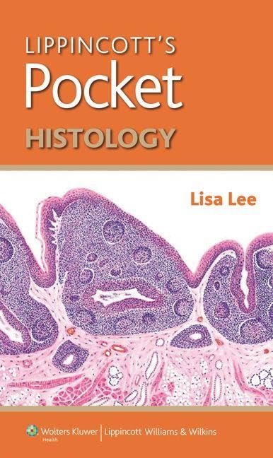 28
28Lippincott's Pocket Histology (Lippincott's Pocket Series)
Lippincott's Pocket Histology is a go-to reference, review and study tool for histology with a strong focus on high-yield topics and presentation. It presents the essential information needed for course and board exam review in a concise, quick-reference format with tables, full-color images, and bullet-point text. The book contains multiple features identifying the clinical significance of concepts, as well as mnemonics to aid in the retention of facts. An index of terms and color-coded systems organization provide easy access to histological facts. This pocket-sized reference intuits how students typically study for exams and provides highly distilled content in one easily portable source.Features include:An introductory section on the basic principles of histology to provide a concise overview of conceptsClinically significant facts for practical applicationOver 300 full-color photomicrographsTables, diagrams, and a complete index of terms for quick referenceMnemonic memory aidsHistological look-a-likes and tips for distinguishing among them
Cover unavailable
29Handbook of Pathogens and Diseases in Cephalopods
 30
30Student Workbook for Illustrated Dental Embryology, Histology and Anatomy
Master the content from your textbook with this helpful study tool! Corresponding to the chapters in Illustrated Dental Embryology, Histology, and Anatomy, 4th Edition, this workbook helps you understand and apply material with case studies, labeling and terminology exercises, new review questions, tooth-drawing guidelines, and more. It includes 32 removable flashcards for quick, convenient study and review of dental structures and plenty of practice questions and case studies to prepare you for classroom and board exams.
A logical organization allows you to focus on areas in which you may need more practice, with units on: (1) labeling, (2) terminology, (3) tooth structure, (4) review questions, and (5) case-based application.
14 detailed case studies include radiographs, clinical photos, profiles, complaints, health histories, and intraoral examination data, each accompanied by multiple-choice questions, to promote critical thinking skills and prepare you for board examinations.
Glossary exercises include crossword puzzles and word searches for practice and review of key terminology.
Guidelines for Tooth Drawing emphasize fundamental principles in tooth design and include detailed instructions, tips, and dimensions for drawing each permanent tooth.
32 detachable flashcards help you master tooth morphology and tooth numbering, with multiple-angle drawings of a permanent tooth on one side of the flashcard and characteristics of that tooth on the back.
Perforated workbook pages are three-hole-punched so that they easily fit into a binder and can be easily removed and submitted to instructors.
NEW! Clinical identification exercises help prepare you for chairside patient care.
NEW! Review questions provide you more opportunities to master the content and prepare for classroom and board exams.
EXPANDED! Structure identification exercises include additional development, microbiological, and anatomical structures for more practice identifying and labeling various parts or structures.
A logical organization allows you to focus on areas in which you may need more practice, with units on: (1) labeling, (2) terminology, (3) tooth structure, (4) review questions, and (5) case-based application.
14 detailed case studies include radiographs, clinical photos, profiles, complaints, health histories, and intraoral examination data, each accompanied by multiple-choice questions, to promote critical thinking skills and prepare you for board examinations.
Glossary exercises include crossword puzzles and word searches for practice and review of key terminology.
Guidelines for Tooth Drawing emphasize fundamental principles in tooth design and include detailed instructions, tips, and dimensions for drawing each permanent tooth.
32 detachable flashcards help you master tooth morphology and tooth numbering, with multiple-angle drawings of a permanent tooth on one side of the flashcard and characteristics of that tooth on the back.
Perforated workbook pages are three-hole-punched so that they easily fit into a binder and can be easily removed and submitted to instructors.
NEW! Clinical identification exercises help prepare you for chairside patient care.
NEW! Review questions provide you more opportunities to master the content and prepare for classroom and board exams.
EXPANDED! Structure identification exercises include additional development, microbiological, and anatomical structures for more practice identifying and labeling various parts or structures.
 31
31John Kellogg Classics: First Book in Physiology and Hygiene: illustrated
This book is intended to give children a good understanding of the structure and functions of the human body.
Cover unavailable
32BOC Study Guide: Histotechnology Certification Exams
 33
33Histotechnology: A Self-Assessment Workbook
Cover unavailable
34Oral Anatomy, Histology and Embryology
The new edition of this now classic book continues to provide dental students with all of the information required to ensure a complete understanding of oral anatomy, histology and embryology as they relate to dental practice. Authored by experts of international renown, the high-quality images and clearly written text found within this volume - features which strongly characterised the high success of previous editions - make study of these challenging topics as easy as possible.
Presents a unique, integrative approach to oral dental science by covering aspects of gross anatomy, tooth morphology, radiology, oral histology and embryology in one volume
Features approximately 1100 high-quality colour images, one quarter of which are previously unpublished
Contains useful 'clinical application' boxes to clearly show relevance of subject area to routine dental practice
Completely revised section on tooth morphology with improved specimens and digital photography
Contains a new chapter on the effects of ageing to highlight the important changes taking place in the oro-dental tissues in an increasingly ageing population
New design aids learning by the inclusion of clearer diagrams and high quality, larger format digital images
Presents a unique, integrative approach to oral dental science by covering aspects of gross anatomy, tooth morphology, radiology, oral histology and embryology in one volume
Features approximately 1100 high-quality colour images, one quarter of which are previously unpublished
Contains useful 'clinical application' boxes to clearly show relevance of subject area to routine dental practice
Completely revised section on tooth morphology with improved specimens and digital photography
Contains a new chapter on the effects of ageing to highlight the important changes taking place in the oro-dental tissues in an increasingly ageing population
New design aids learning by the inclusion of clearer diagrams and high quality, larger format digital images
 35
35Pathology Made Ridiculously Simple
Cover unavailable
36Netter's Neuroscience Flash Cards
Test your knowledge of neuroscience with Dr. David L. Felten's fun, fast, and full-color Netter's Neuroscience Flash Cards. These portable, updated cards let you quiz yourself on anatomy, pathophysiology, and clinical presentation. They now include imaging content and offer increased clinical correlations and new concept summaries. Illustrations from Netter's Atlas of Neuroscience, 2nd Ed., emphasize the essentials of human neuroscience for a quick, yet in-depth review, complete with labeling, explanations, and color codes that cross-reference the atlas.
Master the structures and clinical points most important to a basic medical neuroscience course.
Use the cards before exams or boards, or throughout clinical rotations, residency, or in practice for a fast review of the nervous system.
Cross-reference with Netter's Atlas of Neuroscience, 2nd Ed. for further information on any topic.
Review clinical 'pearls' and helpful summary information on the back of each card to understand the clinical implications of neuroscience concepts.
Use the card set's pre-punched holes and convenient binding ring to carry selected groups of flash cards with you anywhere.
View more neuroimaging examples to assess your grasp of this important subject.
Make clinically important correlations in neuroanatomy, cell biology, and neurophysiology.
Master the structures and clinical points most important to a basic medical neuroscience course.
Use the cards before exams or boards, or throughout clinical rotations, residency, or in practice for a fast review of the nervous system.
Cross-reference with Netter's Atlas of Neuroscience, 2nd Ed. for further information on any topic.
Review clinical 'pearls' and helpful summary information on the back of each card to understand the clinical implications of neuroscience concepts.
Use the card set's pre-punched holes and convenient binding ring to carry selected groups of flash cards with you anywhere.
View more neuroimaging examples to assess your grasp of this important subject.
Make clinically important correlations in neuroanatomy, cell biology, and neurophysiology.
 37
37Anatomy, Histology, & Cell Biology: Pretest Self-Assessment & Review, Fourth Edition
Publisher's Note: Products purchased from Third Party sellers are not guaranteed by the publisher for quality, authenticity, or access to any online entitlements included with the product.
PreTest is the closest you can get to seeing the USMLE Step 1 before you take it
500 USMLE-type questions and answers!
"This edition of PreTest is full of extremely high-yield information in a presentation that is logical and effective. The questions and explanations are invaluable, and the HY tables and figures make it easy to review important material efficiently." -- Gustaf Van Acker III, Fourth Year MD/PhD Candidate, University of Kansas School of Medicine
"This book was an excellent refresher for anyone looking to review information for either their final course exam or for the USMLE Step 1." -- Ben Chidester, Second Year Medical Student, Eastern Virginia Medical School
Great for course review and the USMLE Step 1, Anatomy, Histology, & Cell Biology: PreTest asks the right questions so you'll know the right answers. You'll find 500 clinical-vignette style questions and answers along with complete explanations of correct and incorrect answers. The content has been reviewed by students who recently passed their exams, so you know you are studying the most relevant and up-to-date material possible. No other study guide targets what you really need to know in order to pass like PreTest!
Content that covers all the must-know topics:
High-Yield Facts, Embryology: Early and General, Cell Biology: Membranes, Cell Biology: Cytoplasm, Cell Biology: Intracellular Trafficking, Cell Biology: Nucleus, Epithelium, Connective Tissues, Specialized Connective Tissues: Bones and Cartilage, Muscle and Cell Motility, Nervous System, Cardiovascular System, Blood and Bone Marrow, Lymphoid System and Cellular Immunology, Respiratory System, Integumentary System, Gastrointestinal Tract and Glands, Endocrine Glands, Reproductive System, Urinary System, Eye and Ear, Head and Neck Thorax, Abdomen, Pelvis, Extremities and Spine
PreTest is the closest you can get to seeing the USMLE Step 1 before you take it
500 USMLE-type questions and answers!
"This edition of PreTest is full of extremely high-yield information in a presentation that is logical and effective. The questions and explanations are invaluable, and the HY tables and figures make it easy to review important material efficiently." -- Gustaf Van Acker III, Fourth Year MD/PhD Candidate, University of Kansas School of Medicine
"This book was an excellent refresher for anyone looking to review information for either their final course exam or for the USMLE Step 1." -- Ben Chidester, Second Year Medical Student, Eastern Virginia Medical School
Great for course review and the USMLE Step 1, Anatomy, Histology, & Cell Biology: PreTest asks the right questions so you'll know the right answers. You'll find 500 clinical-vignette style questions and answers along with complete explanations of correct and incorrect answers. The content has been reviewed by students who recently passed their exams, so you know you are studying the most relevant and up-to-date material possible. No other study guide targets what you really need to know in order to pass like PreTest!
Content that covers all the must-know topics:
High-Yield Facts, Embryology: Early and General, Cell Biology: Membranes, Cell Biology: Cytoplasm, Cell Biology: Intracellular Trafficking, Cell Biology: Nucleus, Epithelium, Connective Tissues, Specialized Connective Tissues: Bones and Cartilage, Muscle and Cell Motility, Nervous System, Cardiovascular System, Blood and Bone Marrow, Lymphoid System and Cellular Immunology, Respiratory System, Integumentary System, Gastrointestinal Tract and Glands, Endocrine Glands, Reproductive System, Urinary System, Eye and Ear, Head and Neck Thorax, Abdomen, Pelvis, Extremities and Spine
Cover unavailable
38Color Atlas of Cytology, Histology, and Microscopic Anatomy
A proven classic.
This timeless pocket atlas is the ideal visual companion to histology and cytology textbooks. First published in 1950 and translated into eight languages, Kuehnel's Pocket Atlas of Cytology, Histology and Microscopic Anatomy is a proven classic.
The fully revised and updated fourth edition contains 745 full-color illustrations - almost 200 more than were included in the third edition. Superb, high-quality microphotographs and pathologic stains are accompanied by legends, informative texts, and numerous cross-references.
Key features of the updated fourth edition:
More than 700 high-quality illustrations using advanced techniques in histology and electron microscopy
Practical, information
Concise and focused text
Key concepts and ideas illustrated in less than 550 pages
Ideal for exam preparation, this world-class book is an indispensable visual study tool for medical, dental and biology students. It can also serve as an outstanding review and refresher text.
This timeless pocket atlas is the ideal visual companion to histology and cytology textbooks. First published in 1950 and translated into eight languages, Kuehnel's Pocket Atlas of Cytology, Histology and Microscopic Anatomy is a proven classic.
The fully revised and updated fourth edition contains 745 full-color illustrations - almost 200 more than were included in the third edition. Superb, high-quality microphotographs and pathologic stains are accompanied by legends, informative texts, and numerous cross-references.
Key features of the updated fourth edition:
More than 700 high-quality illustrations using advanced techniques in histology and electron microscopy
Practical, information
Concise and focused text
Key concepts and ideas illustrated in less than 550 pages
Ideal for exam preparation, this world-class book is an indispensable visual study tool for medical, dental and biology students. It can also serve as an outstanding review and refresher text.
Cover unavailable
39Illustrated Dental Embryology, Histology, and Anatomy
Featuring a full-color review of dental structures, Illustrated Dental Embryology, Histology, and Anatomy, 4th Edition provides a complete look at the development, cellular makeup, and morphology of the teeth and associated structures. A clear, reader-friendly writing style makes it easy to understand both basic science and clinical applications, putting the material into the context of everyday dental practice. New to this edition are updates on caries risk, safe levels of fluoride use, and prevention of periodontal disease. Expert authors Margaret Fehrenbach and Tracy Popowics provide an essential background in oral biology for dental hygiene and dental assisting students, including excellent preparation for board exams.
Comprehensive coverage includes all the content needed for an introduction to the developmental, histological, and anatomical foundations of oral health.
Hundreds of full-color anatomical illustrations and clinical and microscopic photographs accompany text descriptions of anatomy and biology.
An approachable writing style covers the latest evidence-based information and makes it easy to grasp and learn to apply the material.
A logical organization separates the book into four units for easier understanding: (1) an introduction to dental structures, (2) dental embryology, (3) dental histology, and (4) dental anatomy.
Key terms open each chapter, accompanied by phonetic pronunciations, and are highlighted within the text, and a glossary provides a quick and handy review and research tool.
Clinical Considerations boxes relate abstract-seeming biological concepts to everyday clinical practice.
Learning outcomes at the beginning of each chapter clearly identify the information you are expected to absorb.
Summary tables and boxes provide quick, easy-to-read summaries of concepts and procedures and serve as useful review and study tools.
Student resources on the Evolve companion website enhance learning with practice quizzes, samplecase studies, review questions, and interactive exercises.
A student workbook offers a wealth of interactive exercises, including labeling/structure identification to master anatomy, word-search and crossword puzzles for vocabulary practice, detailed guidelines for tooth drawing, and illustrated case studies with follow-up questions; in the back of the book, 32 removable flashcards provide practice on identifying permanent teeth and their features and characteristics. Sold separately.
A bibliography lists resource citations for further research and study.
Expert author Margaret Fehrenbach is one of the most trusted names in dental hygiene education, and writes extensively, lectures widely, and consults for many of the major dental manufacturers and supply companies.
NEW! Updated coverage includes the newest evidence-based information on orofacial embryology, especially enamel formation; orofacial histology including fibroblasts, microplicae, keratin, collagen proteins, aging, repair, 3-D tissue engineering, mucoperiosteum, dental pulp stem cells, and platelet-rich plasma; root anatomy; and the latest guidelines on dental biofilm, fluoride use, smile design, periodontal procedures, endoscopy, saliva testing, enamel remineralization, periimplant disease, myofunctional therapy, and orthodontic therapy intervention.
NEW color illustrations, photomicrographs, and diagrams add detail and help to build comprehension.
NEW co-author Tracy Popowics, PhD, provides research and expertise related to advanced dental content.
Comprehensive coverage includes all the content needed for an introduction to the developmental, histological, and anatomical foundations of oral health.
Hundreds of full-color anatomical illustrations and clinical and microscopic photographs accompany text descriptions of anatomy and biology.
An approachable writing style covers the latest evidence-based information and makes it easy to grasp and learn to apply the material.
A logical organization separates the book into four units for easier understanding: (1) an introduction to dental structures, (2) dental embryology, (3) dental histology, and (4) dental anatomy.
Key terms open each chapter, accompanied by phonetic pronunciations, and are highlighted within the text, and a glossary provides a quick and handy review and research tool.
Clinical Considerations boxes relate abstract-seeming biological concepts to everyday clinical practice.
Learning outcomes at the beginning of each chapter clearly identify the information you are expected to absorb.
Summary tables and boxes provide quick, easy-to-read summaries of concepts and procedures and serve as useful review and study tools.
Student resources on the Evolve companion website enhance learning with practice quizzes, samplecase studies, review questions, and interactive exercises.
A student workbook offers a wealth of interactive exercises, including labeling/structure identification to master anatomy, word-search and crossword puzzles for vocabulary practice, detailed guidelines for tooth drawing, and illustrated case studies with follow-up questions; in the back of the book, 32 removable flashcards provide practice on identifying permanent teeth and their features and characteristics. Sold separately.
A bibliography lists resource citations for further research and study.
Expert author Margaret Fehrenbach is one of the most trusted names in dental hygiene education, and writes extensively, lectures widely, and consults for many of the major dental manufacturers and supply companies.
NEW! Updated coverage includes the newest evidence-based information on orofacial embryology, especially enamel formation; orofacial histology including fibroblasts, microplicae, keratin, collagen proteins, aging, repair, 3-D tissue engineering, mucoperiosteum, dental pulp stem cells, and platelet-rich plasma; root anatomy; and the latest guidelines on dental biofilm, fluoride use, smile design, periodontal procedures, endoscopy, saliva testing, enamel remineralization, periimplant disease, myofunctional therapy, and orthodontic therapy intervention.
NEW color illustrations, photomicrographs, and diagrams add detail and help to build comprehension.
NEW co-author Tracy Popowics, PhD, provides research and expertise related to advanced dental content.
 40
40Atlas of Histology with Functional and Clinical Correlations
A comprehensive histology atlas with EXTRAS!
The unique Atlas of Histology with Functional and Clinical Correlations covers fundamental histology topics, integrates this essential information with clinical considerations, and provides multiple opportunities for student review. Explanatory text in each chapter is combined with expanded figure legends in an innovative, integrated layout. The "extras" include:
Key Concepts sections providing comprehensive introductions to and summaries of the chapter in an easy-to-read format.
Over 100 Clinical Correlations that conclude the examination of most normal tissues and structures with a discussion and visual example of relevant pathology.
Numerous Clinical Content boxes highlighted in blue among the running text, sharpening the clinical focus.
Synopsis boxes of the key structural and functional characteristics of cells, tissues, and organs, offering more than 50 opportunities for efficient review.
Full labels directly on the more than 1,300 illustrations and micrographs allowing students to quickly identify structures.
Expanded figure legends reviewing key concepts as applied directly to histology images.
Chapter Summary tables summarizing each chapter or section in a high-yield format.
An Illustrated Glossary of Histological and Pathological Terms introduces the book and the key terms used throughout.
An Appendix explaining the general concept of tissue preparation and staining -- key knowledge for the histology student.
The unique Atlas of Histology with Functional and Clinical Correlations covers fundamental histology topics, integrates this essential information with clinical considerations, and provides multiple opportunities for student review. Explanatory text in each chapter is combined with expanded figure legends in an innovative, integrated layout. The "extras" include:
Key Concepts sections providing comprehensive introductions to and summaries of the chapter in an easy-to-read format.
Over 100 Clinical Correlations that conclude the examination of most normal tissues and structures with a discussion and visual example of relevant pathology.
Numerous Clinical Content boxes highlighted in blue among the running text, sharpening the clinical focus.
Synopsis boxes of the key structural and functional characteristics of cells, tissues, and organs, offering more than 50 opportunities for efficient review.
Full labels directly on the more than 1,300 illustrations and micrographs allowing students to quickly identify structures.
Expanded figure legends reviewing key concepts as applied directly to histology images.
Chapter Summary tables summarizing each chapter or section in a high-yield format.
An Illustrated Glossary of Histological and Pathological Terms introduces the book and the key terms used throughout.
An Appendix explaining the general concept of tissue preparation and staining -- key knowledge for the histology student.
Cover unavailable
41Histology and Cell Biology: An Introduction to Pathology
A vivid, visual tour of a complex and fascinating subject! This book uses a wealth of vivid, full-color images-rather than dense text-to help you master all the histology and cell biology information you need to prepare for your course exams as well as the USMLET Step 1. Clinical correlations drive home the relevance of the material to clinical practice, and new Essential Concepts sections at the end of every chapter facilitate review. And now, online access via STUDENT CONSULT makes this an even more effective learning resource! First prize, Basic and Clinical Sciences category, BMA Awards 2007!
Uses a unique pictorial approach, involving approximately 1,000 full-color illustrations, to make complex principles exceptionally easy to understand and remember. Correlates normal histology with cellular and molecular biology, pathology, and clinical medicine, continually demonstrating the clinical relevance of the material.
Offers new Essential Concepts sections at the end of every chapter to make studying easier than ever.
Includes STUDENT CONSULT access at no additional charge, enabling you to consult the book online, anywhere you go - perform quick searches - add your own notes and bookmarks - follow Integration Links to related bonus content from other STUDENT CONSULT titles-to help you see the connections between diverse disciplines - and more!
Uses a unique pictorial approach, involving approximately 1,000 full-color illustrations, to make complex principles exceptionally easy to understand and remember. Correlates normal histology with cellular and molecular biology, pathology, and clinical medicine, continually demonstrating the clinical relevance of the material.
Offers new Essential Concepts sections at the end of every chapter to make studying easier than ever.
Includes STUDENT CONSULT access at no additional charge, enabling you to consult the book online, anywhere you go - perform quick searches - add your own notes and bookmarks - follow Integration Links to related bonus content from other STUDENT CONSULT titles-to help you see the connections between diverse disciplines - and more!
Cover unavailable
42Intermittent Fasting for Women: How to Lose Weight, Boost Metabolism and Get Healthy
Sick and tired of those fad diets and weight loss programs that never seem to pan out? Seeking a lifestyle change rather than yet another dieting gimmick? Then you should look no further than intermittent fasting. Because far from another useless dieting craze, intermittent fasting is based on centuries of research and harnesses the mechanics of your own physiology so you can burn fat like never before!
Fasting naturally causes our body to switch gears and burn fat deposits rather than the standard sugar and carbs that it normally consumes. And there’s no need to starve yourself: the “intermittent” nature of the fast comes into play on the following day when you end your fast and switch back to your normal eating routine. No reason to quit carbs or count calories. The normal, non-fast day provides a cushion that keeps the body’s metabolism from slowing down.
The problem with so-called starvation diets, is that they will invariably slow down the body’s metabolism as the person’s physiology struggles to cope with the perceived lack of nutrients. And as anyone trying to lose weight knows—this can leave you feeling hungry and exhausted. But intermittent fasting keeps your body’s metabolic engines running on all cylinders.
During an intermittent fasting routine, your body is specifically prepped and primed to burn fat stores one day and then switch back to burning normal food the next, so that your metabolism remains at full steam. If you need to lose weight, boost your metabolism, and get healthy fast—then this guide (written specifically for women) will teach you the basics so you can get started with your own intermittent fast!
In this book you will learn how to:
Implement tested strategies of intermittent fasting Stock up on healthy foods for your fast and non-fast days Create unique meal plans for your weekly fasting routine Reduce stress and anxiety Put your body in detox Prevent the onset of illness Live a healthier lifestyle Improve longevity Raise metabolismBurn fat/lose weight And a whole more!Scroll up and grab your copy so you can start seeing results today!
 43
43Histological and Histochemical Methods, fifth edition: Theory and Practice
This fifth edition of Histological and Histochemical Methods continues to provide a clear and consistent introduction to the techniques, description and analysis of the chemical and physical principles of fixation, tissue processing, staining, enzyme location, immunohistochemistry and other key procedures.
The overall structure of the book remains unchanged, but the content has been heavily revised to update the techniques used in line with recent technological advances. Additionally, there are new sections on:
Artefacts and troubleshooting
Methods for microorganisms and fungi in sections
Methods for various pigments and mineral deposits in tissues
Methods for skeletal elements (bone, cartilage) in whole-mounts
Histological and Histochemical Methods 5e is essential reading for students, lecturers, researchers and professionals using histological and histochemical techniques.
Review of 4th edition: ‘In my opinion this should be a compulsory text in every Histology laboratory.’ Amazon reviewer
The overall structure of the book remains unchanged, but the content has been heavily revised to update the techniques used in line with recent technological advances. Additionally, there are new sections on:
Artefacts and troubleshooting
Methods for microorganisms and fungi in sections
Methods for various pigments and mineral deposits in tissues
Methods for skeletal elements (bone, cartilage) in whole-mounts
Histological and Histochemical Methods 5e is essential reading for students, lecturers, researchers and professionals using histological and histochemical techniques.
Review of 4th edition: ‘In my opinion this should be a compulsory text in every Histology laboratory.’ Amazon reviewer
Cover unavailable
44Wheater's Basic Pathology: A Text, Atlas and Review of Histopathology
Wheater's Basic Pathology: A Text, Atlas and Review of Histopathology, 5th Edition, by Barbara Young, BSc, Med Sci(Hons), PhD, MB, BChir, MRCP, FRCPA, Geraldine O'Dowd, BSc(Hons), MBChB(Hons), FRCPath and William Stewart, BSc (Hons), MBChB, PhD, DipFMS, FRCPath is a pathology resource that offers a comprehensive introduction to the subject first by covering fundamental pathological processes and then addressing the common diseases encountered in systems pathology. Hundreds of high-quality images illustrate the essential features of pathology and make it easy to make definitive comparisons to your own lab samples, while concise captions enable you to quickly and easily understand key points. New online access via Student Consult further enhances your study of the material with additional valuable content including USMLE-style questions and much more. Wheater's Basic Pathology is an excellent companion resource for users of Wheater's Functional Histology, Wheater's Review of Histology, and Basic Pathology, or Robbins and Cotran Pathology Flash Cards.
Offers discussions of basic pathological processes as well as coverage of common diseases encountered in systems pathology for a complete review.
Includes comprehensive updates, with relevant molecular pathology issues explained in the context of the clinical presentation, the treatment implications, and the underlying pathological processes
Presents nearly 650 images of the highest quality that vividly and clearly illustrate the essential features of pathology and enable you to make definitive comparisons to your own lab views.
Features concise text that distills basic and complex information into a coherent explanation to help you understand key points quickly and easily.
Provides keys to the lettering in images at the bottom of each page, saving you time in reviewing figures.
Serves as a companion text to Wheater's Functional Histology, 5th Edition, offering a comparison of normal histology with the pathological changes in disease.
Features a short review section at the end of each chapter covering the key point of the chapter in brief form for the perfect pairing of reference and review.
Includes online access to Student Consult where you'll find the complete text and illustrations from the book, fully searchable . and additional USMLE-style questions for self assessment
Provides additional colored text boxes with clinical-pathological correlations that explain the relevance of the pathological processes underlying common diseases and their complications. FOR FACULTY
Features online access to Evolve Resources including a high-resolution image bank with all the illustrations, figures and tables from the book, and a test bank.
Offers discussions of basic pathological processes as well as coverage of common diseases encountered in systems pathology for a complete review.
Includes comprehensive updates, with relevant molecular pathology issues explained in the context of the clinical presentation, the treatment implications, and the underlying pathological processes
Presents nearly 650 images of the highest quality that vividly and clearly illustrate the essential features of pathology and enable you to make definitive comparisons to your own lab views.
Features concise text that distills basic and complex information into a coherent explanation to help you understand key points quickly and easily.
Provides keys to the lettering in images at the bottom of each page, saving you time in reviewing figures.
Serves as a companion text to Wheater's Functional Histology, 5th Edition, offering a comparison of normal histology with the pathological changes in disease.
Features a short review section at the end of each chapter covering the key point of the chapter in brief form for the perfect pairing of reference and review.
Includes online access to Student Consult where you'll find the complete text and illustrations from the book, fully searchable . and additional USMLE-style questions for self assessment
Provides additional colored text boxes with clinical-pathological correlations that explain the relevance of the pathological processes underlying common diseases and their complications. FOR FACULTY
Features online access to Evolve Resources including a high-resolution image bank with all the illustrations, figures and tables from the book, and a test bank.
 45
45Color Atlas of Veterinary Histology
Blackwell Publishing has acquired 85 titles from LWW's veterinary medicine book list. To place an order or get additional product information go to www.blackwellpublishing.com/vet.
This updated edition offers some significant revisions designed to increase its usefulness for veterinary and veterinary technician students. These include: expanded introductory text for each chapter; expanded legends on a selective basis throughout the book; standardized terms to conform with the Nomina Anatomica Veterinaria, 1996 Edition; views at higher magnification; twenty new color figures; detailed Glossary of Terms.
This updated edition offers some significant revisions designed to increase its usefulness for veterinary and veterinary technician students. These include: expanded introductory text for each chapter; expanded legends on a selective basis throughout the book; standardized terms to conform with the Nomina Anatomica Veterinaria, 1996 Edition; views at higher magnification; twenty new color figures; detailed Glossary of Terms.
 46
46Functional Histology
The study of histology requires that students be able to recognise structures within cells and tissues at varying levels of magnification, and understand their function within the human body. Since histology is a visual subject, high quality images of cells and tissues are vital as a component of course material. In this book, concise text relates the structures seen in the images to biological function, and integrates clinical relevance by describing how the histology of tissues is affected in abnormal conditions.
WINNER - Best Designed Tertiary and Further Education Book at the "Australian 58th Annual Book Design Awards," 20th May 2010
WINNER - Best Designed Tertiary and Further Education Book at the "Australian 58th Annual Book Design Awards," 20th May 2010
Cover unavailable
47Or Perish in the Attempt: The Hardship and Medicine of the Lewis and Clark Expedition
David J. Peck’s Or Perish in the Attempt ingeniously combines the remarkable adventures of Lewis and Clark with an examination of the health problems their expedition faced. Formidable problems indeed, but the author patiently, expertly—and humorously—guides us through the medical travails of the famous journey, juxtaposing treatment then against remedy now. The result is a fascinating book that sheds new light not only on Lewis and Clark and the men and one remarkable woman (and her infant) who accompanied them along an eight-thousand-mile wilderness path but also on the practice of medicine in their time and place.
Cover unavailable
48Color Atlas of Histology
This compact atlas provides medical, dental, allied health, and biology students with an outstanding collection of histology images for all of the major tissue classes and body systems. The Fourth Edition features a completely re-digitized image collection, along with other hallmarks of this atlas that have been proven over time, including its concise writing style, compact size/format, and quick introduction/background for each chapter that reviews essential concepts, histophysiology, and clinical considerations.
A bound-in interactive student CD-ROM contains all the images from the book with compare and full-screen view modes, zoom, label rollover and on/off features, along with USMLE-style and identification review questions.
A bound-in interactive student CD-ROM contains all the images from the book with compare and full-screen view modes, zoom, label rollover and on/off features, along with USMLE-style and identification review questions.
Cover unavailable
49Basic Histology Flash Cards
Flashcards derived from one of the most popular histology texts ever published.
Cover unavailable
50Histology and Cell Biology: Examination and Board Review, Fifth Edition
Publisher's Note: Products purchased from Third Party sellers are not guaranteed by the publisher for quality, authenticity, or access to any online entitlements included with the product.
A complete one-stop review of the clinically important aspects of histology and cell biology--user-friendly, concise, and packed with learning aids!
The ideal review for course exams and the USMLE!
4 STAR DOODY'S REVIEW!
"This is a wonderful resource for students of medicine, dentistry, and the allied health sciences. The book combines traditional topics in histology with elements of modern cell biology and medical physiology.... This is the body of information that students of microscopic anatomy need to know to understand the foundations of clinical medicine and succeed on future licensing examinations. Students will use this book to review key concepts in modern histology."--Doody's Review Service
This popular title in the LANGE series is specifically designed to help you make the most of your study time--whether you're studying histology and cell biology for the first time or reviewing for course exams or the USMLE. With this focused review you will be able to pinpoint your weak areas, and then improve your comprehension with learning aids especially designed to help you understand and retain even the most difficult material.
You will find complete easy-to-follow coverage of all the need-to-know material: fundamental concepts, the four basic tissues types, and organs and organ systems--presented in a consistent, time-saving design. At the conclusion of the book, you will find a Diagnostic Final Exam that has been updated with longer, case-related stems that mimic the USMLE Step 1 examination.
Each chapter is devoted to one specific topic and includes learning aids such as:
Objectives that point out significant facts and concepts that you must know about each topic
Max Yield(TM) study questions that direct you to key facts needed to master material most often covered on exams
A synopsis presented in outline form that reviews all the basic histology and related cell biology covered on exams
Multiple-choice questions written in a style most commonly used in medical school
NEW to this Edition:
Thoroughly revised Q&A
Completely updated text and practice questions to reflect current knowledge
Information added to each chapter regarding relevant pathology/clinical issues; possibly as a separate colored box
Visit www.LangeTextbooks.com to access valuable resources and study aids.
Thorough coverage you won't find anywhere else!
FUNDAMENTAL CONCEPTS: Methods of Study, The Plasma Membrane & Cytoplasm, The Nucleus & Cell Cycle, THE FOUR BASIC TISSUE TYPES: Epithelial Tissue, Connective Tissue, Adipose Tissue, Cartilage, Bone, Integrative Multiple-Choice Questions: Connective Tissues Nerve Tissue, Muscle Tissue, Integrative Multiple-Choice Questions: Basic Tissue Types, ORGANS & ORGAN SYSTEMS: Circulatory System, Peripheral Blood, Hematopoiesis, Lymphoid System, Digestive Tract, Glands Associated with the Digestive Tract, Integrative Multiple-Choice Questions: Digestive System, Respiratory System, Skin, Urinary System, Pituitary & Hypothalamus, Adrenals, Islets of Langerhans, Thyroid, Parathyroids, & Pineal Body, Male Reproductive System, Female Reproductive System, Integrative Multiple-Choice Questions: Endocrine System, Sense Organs, Diagnostic Final Examination
A complete one-stop review of the clinically important aspects of histology and cell biology--user-friendly, concise, and packed with learning aids!
The ideal review for course exams and the USMLE!
4 STAR DOODY'S REVIEW!
"This is a wonderful resource for students of medicine, dentistry, and the allied health sciences. The book combines traditional topics in histology with elements of modern cell biology and medical physiology.... This is the body of information that students of microscopic anatomy need to know to understand the foundations of clinical medicine and succeed on future licensing examinations. Students will use this book to review key concepts in modern histology."--Doody's Review Service
This popular title in the LANGE series is specifically designed to help you make the most of your study time--whether you're studying histology and cell biology for the first time or reviewing for course exams or the USMLE. With this focused review you will be able to pinpoint your weak areas, and then improve your comprehension with learning aids especially designed to help you understand and retain even the most difficult material.
You will find complete easy-to-follow coverage of all the need-to-know material: fundamental concepts, the four basic tissues types, and organs and organ systems--presented in a consistent, time-saving design. At the conclusion of the book, you will find a Diagnostic Final Exam that has been updated with longer, case-related stems that mimic the USMLE Step 1 examination.
Each chapter is devoted to one specific topic and includes learning aids such as:
Objectives that point out significant facts and concepts that you must know about each topic
Max Yield(TM) study questions that direct you to key facts needed to master material most often covered on exams
A synopsis presented in outline form that reviews all the basic histology and related cell biology covered on exams
Multiple-choice questions written in a style most commonly used in medical school
NEW to this Edition:
Thoroughly revised Q&A
Completely updated text and practice questions to reflect current knowledge
Information added to each chapter regarding relevant pathology/clinical issues; possibly as a separate colored box
Visit www.LangeTextbooks.com to access valuable resources and study aids.
Thorough coverage you won't find anywhere else!
FUNDAMENTAL CONCEPTS: Methods of Study, The Plasma Membrane & Cytoplasm, The Nucleus & Cell Cycle, THE FOUR BASIC TISSUE TYPES: Epithelial Tissue, Connective Tissue, Adipose Tissue, Cartilage, Bone, Integrative Multiple-Choice Questions: Connective Tissues Nerve Tissue, Muscle Tissue, Integrative Multiple-Choice Questions: Basic Tissue Types, ORGANS & ORGAN SYSTEMS: Circulatory System, Peripheral Blood, Hematopoiesis, Lymphoid System, Digestive Tract, Glands Associated with the Digestive Tract, Integrative Multiple-Choice Questions: Digestive System, Respiratory System, Skin, Urinary System, Pituitary & Hypothalamus, Adrenals, Islets of Langerhans, Thyroid, Parathyroids, & Pineal Body, Male Reproductive System, Female Reproductive System, Integrative Multiple-Choice Questions: Endocrine System, Sense Organs, Diagnostic Final Examination
 51
51Dellmann's Textbook of Veterinary Histology, with CD
The leading veterinary histology text returns with a fully updated sixth edition. Written in a concise, easy-to-understand that's a pleasure to read, this new edition continues the student-friendly tradition originated by Dr. Dellman, presenting the basics of histology including cytology and microscopic anatomy. The Sixth Edition focuses on the most current knowledge of cell, tissue and organ structure and function. All information has been fully revised and updated by the authors, both experts in their fields. Written with first year veterinary students in mind, it is also an important resource for veterinarians, graduate students, and others who require information on animal tissue structure and function.
Highlights of the Sixth Edition include:
New images and line drawings have been added to enhance the student's understanding of concepts. Two-page insert contains full-color histology images. Comprehensive listings of suggested readings at the end of each chapter encourage further study. The text is organized by body region, allowing the presentation to emphasize comparative species information so students can better appreciate how species differ in regard to key structures. Whether you're a veterinary student or practicing professional, you should have this classic histology reference as part of your working library.
Highlights of the Sixth Edition include:
New images and line drawings have been added to enhance the student's understanding of concepts. Two-page insert contains full-color histology images. Comprehensive listings of suggested readings at the end of each chapter encourage further study. The text is organized by body region, allowing the presentation to emphasize comparative species information so students can better appreciate how species differ in regard to key structures. Whether you're a veterinary student or practicing professional, you should have this classic histology reference as part of your working library.
 52
52Histology for Pathologists
Now completely revised and updated, this ground-breaking text focuses on the borderland between histology and pathology. The text describes human histology and its variations, with emphasis on avoiding overdiagnosis or misdiagnosis of normal variants. Presented by leading international specialists in thology,histopathology, anatomical pathology, surgical pathology, and neuropathology, the text is innovative, well organized and generously illustrated. Coverage includes the structure, composition, and function of tissues from a pathologic perspective; a systematic review of each major organ system, as well as structures of special significance; and histologic subtleties, such as unusual variations in staining reactions, little-known fixation artifacts, and frequently missed gross observations.
This edition is reorganized to better reflect an organ-system approach to histology. Immunohistochemical findings in normal tissues have been added, based on the latest antibody panels.
A bound-in CD-ROM image bank is included.
This edition is reorganized to better reflect an organ-system approach to histology. Immunohistochemical findings in normal tissues have been added, based on the latest antibody panels.
A bound-in CD-ROM image bank is included.
Cover unavailable
53Clinical Anatomy Of The Visual System
This concise source for the clinical anatomy of the visual system covers the clinical anatomy of the eye, its adnexa, and the visual pathways in a well-illustrated, well-referenced format. It balances histologic content of the microscopic anatomy with functional aspects of the eye and visual system. Clear illustrations complement the text and provide clinical information on diseases and disorders that represent departures from normal clinical anatomy.
Full color illustrations throughout enhance the anatomical and clinical information.
The only anatomy text written by an optometrist for optometrists and students - it provides a strong foundation for recognizing and understanding clinical situations, problems, and treatments.
Clinical Comment sections have been expanded to include more clinical correlation comments and put knowledge of ocular anatomy into a practice perspective.
Clinical appearance of normal structures is now included, with normal photos of the cornea, iris, lens, retina, etc., to show how appearance is directly related to structure.
New information on aging changes in ocular tissue and the visual pathway keeps readers current with the expanding field of geriatric care and the changes that occur to normal anatomy.
Full color illustrations throughout enhance the anatomical and clinical information.
The only anatomy text written by an optometrist for optometrists and students - it provides a strong foundation for recognizing and understanding clinical situations, problems, and treatments.
Clinical Comment sections have been expanded to include more clinical correlation comments and put knowledge of ocular anatomy into a practice perspective.
Clinical appearance of normal structures is now included, with normal photos of the cornea, iris, lens, retina, etc., to show how appearance is directly related to structure.
New information on aging changes in ocular tissue and the visual pathway keeps readers current with the expanding field of geriatric care and the changes that occur to normal anatomy.
Cover unavailable
54Sushi: Food for the Eye, the Body & the Soul
"It is clear that serious research, as well as much imagination, went into every page. It has become my new 'go-to' bible when I need a shot of inspiration."
Ken Oringer, internationally renowned and award-winning chef
Clio Restaurant, Uni Sashimi Bar, Boston
"Congratulations on writing such an aesthetically beautiful, informative and inspiring book. ... I shall not hesitate to recommend your book to those colleagues, who like me, are fascinated by Sushi and who will surely be captivated, like me, turning every page."
Dr. Ian C. Forster, April, 2011
- - -
In recent decades, sushi has gone from being a rather exotic dish, eaten by relatively few outside of Japan, to a regular meal for many across the world. It is quickly gathering the attention of chefs and nutritionists everywhere. It has even made its way into numerous home kitchens where people have patiently honed the specialized craft required to prepare it. Few have been more attuned to this remarkable transition than Ole G. Mouritsen, an esteemed Danish scientist and amateur chef who has had a lifelong fascination with sushi's central role in Japanese culinary culture.
Sushi for the eye, the body, and the soul is a unique melange of a book. In it, Mouritsen discusses the cultural history of sushi then uses his scientific prowess to deconstruct and explain the complex chemistry of its many subtle and sharp taste sensations. He also offers insights from years of honing his own craft as a sushi chef, detailing how to choose and prepare raw ingredients, how to decide which tools and techniques to use, and how to arrange and present various dishes.
Sushi is irresistible for both its simplicity and the hypnotic performance-art aspects that go into its preparation. With clear prose and straightforward instructions, Mouritsen looks at every facet of sushi in a book that is as accessible as it is informative, as useful as it is fun.
Ken Oringer, internationally renowned and award-winning chef
Clio Restaurant, Uni Sashimi Bar, Boston
"Congratulations on writing such an aesthetically beautiful, informative and inspiring book. ... I shall not hesitate to recommend your book to those colleagues, who like me, are fascinated by Sushi and who will surely be captivated, like me, turning every page."
Dr. Ian C. Forster, April, 2011
- - -
In recent decades, sushi has gone from being a rather exotic dish, eaten by relatively few outside of Japan, to a regular meal for many across the world. It is quickly gathering the attention of chefs and nutritionists everywhere. It has even made its way into numerous home kitchens where people have patiently honed the specialized craft required to prepare it. Few have been more attuned to this remarkable transition than Ole G. Mouritsen, an esteemed Danish scientist and amateur chef who has had a lifelong fascination with sushi's central role in Japanese culinary culture.
Sushi for the eye, the body, and the soul is a unique melange of a book. In it, Mouritsen discusses the cultural history of sushi then uses his scientific prowess to deconstruct and explain the complex chemistry of its many subtle and sharp taste sensations. He also offers insights from years of honing his own craft as a sushi chef, detailing how to choose and prepare raw ingredients, how to decide which tools and techniques to use, and how to arrange and present various dishes.
Sushi is irresistible for both its simplicity and the hypnotic performance-art aspects that go into its preparation. With clear prose and straightforward instructions, Mouritsen looks at every facet of sushi in a book that is as accessible as it is informative, as useful as it is fun.
Cover unavailable
55Playing the Ponies and Other Medical Mysteries Solved
With over forty years of experience as a sought after diagnostician, Dr. Stuart Mushlin has cracked his share of medical mysteries, ones in which there are bigger gambles than playing the ponies at the track. Some of his patients show up with puzzling symptoms, calling for savvy medical detective work. Others seem to present cut-and-dry cases, but they turn out to be suffering from rare or serious conditions.
In Playing the Ponies and Other Medical Mysteries Solved, Dr. Mushlin shares some of the most intriguing cases he has encountered, revealing the twists and turns of each patient’s diagnosis and treatment process. Along the way, he imparts the secrets to his success as a medical detective—not specialized high-tech equipment, but time-honored techniques like closely observing, touching, and listening to patients. He also candidly describes cases where he got things wrong, providing readers with honest insights into both the joys and dilemmas of his job.
Dr. Mushlin does not just treat diseases; he treats people. And this is not just a book about the ailments he diagnosed; it is also about the scared, uncertain, ailing individuals he helped in the process. Filled with real-life medical stories you’ll have to read to believe, Playing the Ponies is both a suspenseful page-turner and a heartfelt reflection on a life spent caring for patients.
In Playing the Ponies and Other Medical Mysteries Solved, Dr. Mushlin shares some of the most intriguing cases he has encountered, revealing the twists and turns of each patient’s diagnosis and treatment process. Along the way, he imparts the secrets to his success as a medical detective—not specialized high-tech equipment, but time-honored techniques like closely observing, touching, and listening to patients. He also candidly describes cases where he got things wrong, providing readers with honest insights into both the joys and dilemmas of his job.
Dr. Mushlin does not just treat diseases; he treats people. And this is not just a book about the ailments he diagnosed; it is also about the scared, uncertain, ailing individuals he helped in the process. Filled with real-life medical stories you’ll have to read to believe, Playing the Ponies is both a suspenseful page-turner and a heartfelt reflection on a life spent caring for patients.
Cover unavailable
56Clinical Anatomy and Physiology of the Visual System
Taking the place of the multiple texts traditionally needed to cover visual anatomy and physiology, Clinical Anatomy and Physiology of the Visual System, 3rd Edition dramatically lightens your load by providing one book that covers it all! This concise, well-referenced resource contains information on the clinical anatomy of the eye, its adnexa and visual pathways, histologic information, plus newly added content on physiology of the human ocular structures. Vivid illustrations complement the text and provide clinical information on diseases and disorders that represent departures from normal clinical anatomy.
Comprehensive physiology coverage clarifies the integration between structure and function, eliminating your need for multiple books on the anatomy and physiology of the visual system.
An emphasis on clinical application helps you better understand the processes that occur in disease and dysfunction.
Genetic information keeps you current with the latest developments in visual anatomy and physiology.
Full-color illustrations throughout the text enhance your understanding of anatomical and clinical information.
UNIQUE! Clinical Comment sections provide a solid foundation for recognizing and understanding clinical situations, conditions, diseases, and treatments.
Photos of normal eye structures illustrate clinical appearance and demonstrate how appearance is directly related to structure.
Geriatric coverage, including aging changes in ocular tissue and the visual pathway, keeps you up-to-date with the expanding field of geriatric care.
UNIQUE! Expert coverage written by an actual optometrist gives you a practical framework for recognizing and understanding clinical situations, problems, and treatments.
Comprehensive physiology coverage clarifies the integration between structure and function, eliminating your need for multiple books on the anatomy and physiology of the visual system.
An emphasis on clinical application helps you better understand the processes that occur in disease and dysfunction.
Genetic information keeps you current with the latest developments in visual anatomy and physiology.
Full-color illustrations throughout the text enhance your understanding of anatomical and clinical information.
UNIQUE! Clinical Comment sections provide a solid foundation for recognizing and understanding clinical situations, conditions, diseases, and treatments.
Photos of normal eye structures illustrate clinical appearance and demonstrate how appearance is directly related to structure.
Geriatric coverage, including aging changes in ocular tissue and the visual pathway, keeps you up-to-date with the expanding field of geriatric care.
UNIQUE! Expert coverage written by an actual optometrist gives you a practical framework for recognizing and understanding clinical situations, problems, and treatments.
 57
57Basic Histology: Text & Atlas
Cover unavailable
58Stevens y Lowe. Histología humana + StudentConsult
* La nueva edición de la obra continua con la filosofía habitual, ya que se trata de un texto profusamente ilustrado ( en 448 páginas se contabilizan cerca de 900 imágenes) en el que se presentan los conceptos básicos y fundamentales que el estudiante debe adquirir en el que se incluye correlación clínica y casos de estudio en donde se relaciona estructura y función. • Se trata de un texto muy bien organizado, en el que la estructura de los capítulos es altamente homogénea. Tiene un elevado carácter pedagógico, de forma que el objetivo que persiguen los autores es obtener un texto en donde la información pueda localizarse de forma rápida y fácil, de forma que los conceptos sean fácilmente asimilables. • A lo largo de los capítulos pueden localizarse los siguientes elementos que le confieren este gran valor didáctico: - Cuadros "Advanced Concept": en ellos se profundiza en conceptos explicados o citados brevemente en el texto. - Cuadros de "Clinical exemple": en ellos se explican de forma detallada diferentes patologías, acompañados normalmente por su imagen clínica correspondiente. - Cuadros "Practical Histology ": en los que se enseña al estudiante como se vería el tejido estudiado si en ese momento lo estuviera viendo al microscopio. - Cuadros de "key facts", en los que se listan los conceptos clave del estilo "need to know" - Preguntas de repaso: al final de cada uno de los capítulos se incluye un cuadro en el que se plantean 4 preguntas cortas con 5 opciones de respuesta y se plantea de forma breve un caso clínico sobre el que se hace una pregunta. Las respuestas correspondientes se encuentran al final del libro.
Cover unavailable
59Histologic Preparations: Common Problems and Their Solutions
The "how to" guide to good slide preparation, Histologic Preparations: Common Problems and Their Solutions, was developed by the Histotechnology Committee of the College of American Pathologists (CAP) in conjunction with the National Society for Histotechnology (NSH). Building on data and images from the NSH/CAP histology quality assurance program, HistoQIP, the book presents photographic examples of well-prepared slides followed by numerous examples of associated problems and their solutions. Histologic Preparations is a reference text as well as a teaching tool. Written for pathologists, pathology residents, histotechnologists, and histotechnicians as well as histology students, this thorough book contains troubleshooting techniques for the most common artifacts and problems incurred in routine histologic preparations. Some of the topics covered are fixation and processing; microtomy; frozen sections; H&E and Gram stains; mycobacteria, H pylori, spirochetes, and fungi; and trichrome, reticulin, elastin, basement membrane, mucin, amyloid, and immunohistochemical stains. This timely, practical, and straightforward book will be turned to repeatedly as a reliable reference in the laboratory.
 60
60Cell Biology Bold and Easy Science Coloring: Color cells, tissues and organelles, hand drawn by a science teacher with over 20 years experience of explaining tricky concepts in a clear and simple way.
Coloring Book! Created by an experienced science teacher with over 20 years in the classroom, this book makes the complex world of cell biology accessible, engaging, and fun for all ages—from toddlers to grandparents.Perfect for anyone who loves science or simply wants to learn more about cell structure, this coloring book features 24 unique, hand-drawn images of plant and animal cells, complete with all the essential organelles typically found in each. Whether you're a curious beginner or a biology enthusiast, this book offers a creative way to explore the fundamental building blocks of life.Designed for coloring with pencils, crayons, or markers, the bold, easy-to-follow illustrations make learning stress-free and enjoyable. (Pro tip: When using alcohol-based markers, place a sheet of paper behind each page to prevent bleed-through.)Uncover the wonders of biology one color at a time, and let this coloring book turn a challenging subject into an enjoyable learning experience for all!
 61
61A Practical Guide to the Histology of the Mouse
A Practical Guide to the Histology of the Mouse provides a full-colour atlas of mouse histology. Mouse models of disease are used extensively in biomedical research with many hundreds of new models being generated each year. Complete phenotypic analysis of all of these models can benefit from histologic review of the tissues.
This book is aimed at veterinary and medical pathologists who are unfamiliar with mouse tissues and scientists who wish to evaluate their own mouse models. It provides practical guidance on the collection, sampling and analysis of mouse tissue samples in order to maximize the information that can be gained from these tissues. As well as illustrating the normal microscopic anatomy of the mouse, the book also describes and explains the common anatomic variations, artefacts associated with tissue collection and background lesions to help the scientist to distinguish these changes from experimentally- induced lesions.
This will be an essential bench-side companion for researchers and practitioners looking for an accessible and well-illustrated guide to mouse pathology.
Written by experienced pathologists and specifically tailored to the needs of scientists and histologistsFull colour throughoutProvides advice on sampling tissues, necropsy and recording dataIncludes common anatomic variations, background lesions and artefacts which will help non-experts understand whether histologic variations seen are part of the normal background or related to their experimental manipulation
This book is aimed at veterinary and medical pathologists who are unfamiliar with mouse tissues and scientists who wish to evaluate their own mouse models. It provides practical guidance on the collection, sampling and analysis of mouse tissue samples in order to maximize the information that can be gained from these tissues. As well as illustrating the normal microscopic anatomy of the mouse, the book also describes and explains the common anatomic variations, artefacts associated with tissue collection and background lesions to help the scientist to distinguish these changes from experimentally- induced lesions.
This will be an essential bench-side companion for researchers and practitioners looking for an accessible and well-illustrated guide to mouse pathology.
Written by experienced pathologists and specifically tailored to the needs of scientists and histologistsFull colour throughoutProvides advice on sampling tissues, necropsy and recording dataIncludes common anatomic variations, background lesions and artefacts which will help non-experts understand whether histologic variations seen are part of the normal background or related to their experimental manipulation
 62
62The Extracellular Matrix and Ground Regulation: Basis for a Holistic Biological Medicine
The workings of the suitable environment for cells—called the extracellular matrix (ECM) and ground regulation—has occupied the European medical tradition since the early part of the 20th century. As it has become more clear that the origin of disease and its first signals register in the connective tissue, or myofascia, cellular pathologists and biochemists have sought to circumscribe networks of cell communication and microcirculation in the ECM.
Alfred Pischinger (1899-1982) continued this line of work by further studying, in work published from 1926 through the late seventies, the connections of the ECM to the hormonal and autonomic systems. In the last twenty years Professor and Doctor of Natural Sciences Hartmut Heine and his colleagues have carried on Pischinger’s work, here summarized in one volume. Part One encompasses theoretical underpinnings; Parts Two and Three address applications and directions for further research.
This updated English-language translation not only is an account of the work of Pischinger’s successors—Heine, Otto Bergsmann, and Felix Perger, (the three editors of this volume) and their many colleagues—but notes the positive development of complementary therapies based on this understanding of histology. Acupuncture is referenced directly. Both in Europe and the States the work of manual therapists, including Rolfers, cranio- sacral therapists, and other somatic disciplines have been informed for many years by Pischinger’s outsider model of how changes in the EMC register in the central nervous system and the brain, and are conveyed back to the periphery and connected organs. Heine’s exciting recent work shows that the regulation and construction of the ECM have relationships to cybernetic non-linear systems and phase transitions.
Alfred Pischinger (1899-1982) continued this line of work by further studying, in work published from 1926 through the late seventies, the connections of the ECM to the hormonal and autonomic systems. In the last twenty years Professor and Doctor of Natural Sciences Hartmut Heine and his colleagues have carried on Pischinger’s work, here summarized in one volume. Part One encompasses theoretical underpinnings; Parts Two and Three address applications and directions for further research.
This updated English-language translation not only is an account of the work of Pischinger’s successors—Heine, Otto Bergsmann, and Felix Perger, (the three editors of this volume) and their many colleagues—but notes the positive development of complementary therapies based on this understanding of histology. Acupuncture is referenced directly. Both in Europe and the States the work of manual therapists, including Rolfers, cranio- sacral therapists, and other somatic disciplines have been informed for many years by Pischinger’s outsider model of how changes in the EMC register in the central nervous system and the brain, and are conveyed back to the periphery and connected organs. Heine’s exciting recent work shows that the regulation and construction of the ECM have relationships to cybernetic non-linear systems and phase transitions.
 63
63Textbook of Veterinary Histology
Logically organized by body system, this comprehensive resource provides in-depth coverage of the structure and function of the cells, tissues, and organs of a wide range of domestic animal species. Bridging the gap between the physiology and the gross anatomy of organisms, it also explores new discoveries being made in the areas of molecular biology and cytogenetics.
Full-color coverage throughout, including more than 400 color figures that are grouped into major sections for quick reference
A wealth of electron micrographs and color micrographs demonstrate cell, tissue, and organ structure
A complete art program integrates illustrations and diagrams of cells and tissues to highlight structural-functional correlates
Helpful tables of histological features in each chapter summarize key concepts
A succinct style and format makes it easy to quickly find important information
Chapters begin with a list of key points
The author is a trained morphologist and has taught veterinary histology at the University of Florida College of Veterinary Medicine for more than 15 years
Full-color coverage throughout, including more than 400 color figures that are grouped into major sections for quick reference
A wealth of electron micrographs and color micrographs demonstrate cell, tissue, and organ structure
A complete art program integrates illustrations and diagrams of cells and tissues to highlight structural-functional correlates
Helpful tables of histological features in each chapter summarize key concepts
A succinct style and format makes it easy to quickly find important information
Chapters begin with a list of key points
The author is a trained morphologist and has taught veterinary histology at the University of Florida College of Veterinary Medicine for more than 15 years
 64
64Netter's Essential Histology
"Netter's Essential Histology" integrates gross anatomy and embryology with classic histology slides and cutting-edge scanning electron microscopy to give you a rich visual understanding of this complex subject. This histology textbook-atlas has a strong anatomy foundation and utilizes a variety of visual elements - including Netter illustrations and light and electron micrographs - to teach you the most indispensible histologic concepts and their clinical relevance. Excellent as both a reference and a review, "Netter's Essential Histology "will serve you well at any stage of your healthcare career.
 65
65Procedures in Cosmetic Dermatology Series: Soft Tissue Augmentation
 66
66Forerunners of Mammals: Radiation • Histology • Biology (Life of the Past)
About 320 million years ago a group of reptiles known as the synapsids emerged and forever changed Earth’s ecological landscapes. This book discusses the origin and radiation of the synapsids from their sail-backed pelycosaur ancestor to their diverse descendants, the therapsids or mammal-like reptiles, that eventually gave rise to mammals. It further showcases the remarkable evolutionary history of the synapsids in the Karoo Basin of South Africa and the environments that existed at the time. By highlighting studies of synapsid bone microstructure, it offers a unique perspective of how such studies are utilized to reconstruct various aspects of biology, such as growth dynamics, biomechanical function, and the attainment of sexual and skeletal maturity. A series of chapters outline the radiation and phylogenetic relationships of major synapsid lineages and provide direct insight into how bone histological analyses have led to an appreciation of these enigmatic animals as once-living creatures. The penultimate chapter examines the early radiation of mammals from their nonmammalian cynodont ancestors, and the book concludes by engaging the intriguing question of when and where endothermy evolved among the therapsids.
Cover unavailable
67Delivered by Midwives: African American Midwifery in the Twentieth-Century South
Winner of the 2019 American Association for the History of Nursing Lavinia L. Dock Award for Exemplary Historical Research and Writing in a Book
"Catchin' babies" was merely one aspect of the broad role of African American midwives in the twentieth-century South. Yet, little has been written about the type of care they provided or how midwifery and maternity care evolved under the increasing presence of local and federal health care structures.
Using evidence from nursing, medical, and public health journals of the era; primary sources from state and county departments of health; and personal accounts from varied practitioners, Delivered by Midwives: African American Midwifery in the Twentieth-Century South provides a new perspective on the childbirth experience of African American women and their maternity care providers. Author Jenny M. Luke moves beyond the usual racial dichotomies to expose a more complex shift in childbirth culture, revealing the changing expectations and agency of African American women in their rejection of a two-tier maternity care system and their demands to be part of an inclusive, desegregated society.
Moreover, Luke illuminates valuable aspects of a maternity care model previously discarded in the name of progress. High maternal and infant mortality rates led to the passage of the Sheppard-Towner Maternity and Infancy Protection Act in 1921. This marked the first attempt by the federal government to improve the welfare of mothers and babies. Almost a century later, concern about maternal mortality and persistent racial disparities have forced a reassessment. Elements of the long-abandoned care model are being reincorporated into modern practice, answering current health care dilemmas by heeding lessons from the past.
"Catchin' babies" was merely one aspect of the broad role of African American midwives in the twentieth-century South. Yet, little has been written about the type of care they provided or how midwifery and maternity care evolved under the increasing presence of local and federal health care structures.
Using evidence from nursing, medical, and public health journals of the era; primary sources from state and county departments of health; and personal accounts from varied practitioners, Delivered by Midwives: African American Midwifery in the Twentieth-Century South provides a new perspective on the childbirth experience of African American women and their maternity care providers. Author Jenny M. Luke moves beyond the usual racial dichotomies to expose a more complex shift in childbirth culture, revealing the changing expectations and agency of African American women in their rejection of a two-tier maternity care system and their demands to be part of an inclusive, desegregated society.
Moreover, Luke illuminates valuable aspects of a maternity care model previously discarded in the name of progress. High maternal and infant mortality rates led to the passage of the Sheppard-Towner Maternity and Infancy Protection Act in 1921. This marked the first attempt by the federal government to improve the welfare of mothers and babies. Almost a century later, concern about maternal mortality and persistent racial disparities have forced a reassessment. Elements of the long-abandoned care model are being reincorporated into modern practice, answering current health care dilemmas by heeding lessons from the past.
Cover unavailable
68Atlas of Descriptive Histology
The Atlas of Descriptive Histology is the most comprehensive histology atlas available. It covers all tissues and organs, each subject prefaced by introductory boxed text. Full-color high-resolution micrographs, gathered from colleagues both domestic and international, provide superb image quality. By showing images of the same area at successively increasing magnifications, the book provides students with an experience similar to that they would get from an actual microscope. Careful descriptions of each micrograph include those details a student should be able to recognize.
RESOURCES
For the Student
Companion Website
Each copy of the Atlas of Descriptive Histology includes an access code that gives the student a subscription to the textbook's Companion Website. The site features a flexible image viewer that allows the student to view any of the micrographs in the book. Features of the image viewer include:
*Each micrograph from each plate in the textbook is provided separately.
*A zoom tool allows the student to view the micrograph at either low resolution or high resolution--providing greatly increased detail.
*Labels and leaders can each be displayed or hidden independently.
*A self-quiz mode allows the student to test themselves on structure identification.
*Each micrograph can be downloaded for incorporation into student notes or presentations.
For the Instructor
Instructor's Resource Library
The Atlas of Descriptive Histology Instructor's Resource Library includes multiple versions of every micrograph from every plate in the textbook, as well as an instructor version of the image viewer from the Companion Website. The IRL includes the following:
*Micrographs provided in three formats: Without leaders or labels, with leaders only, and with leaders and labels.
*All images provided as both low-resolution and high-resolution JPEGs.
*A PowerPoint presentation of all images for each chapter.
*The instructor version of the image viewer, which includes all the features described above, with the addition of a pen tool that allows drawing on the image during lecture.
RESOURCES
For the Student
Companion Website
Each copy of the Atlas of Descriptive Histology includes an access code that gives the student a subscription to the textbook's Companion Website. The site features a flexible image viewer that allows the student to view any of the micrographs in the book. Features of the image viewer include:
*Each micrograph from each plate in the textbook is provided separately.
*A zoom tool allows the student to view the micrograph at either low resolution or high resolution--providing greatly increased detail.
*Labels and leaders can each be displayed or hidden independently.
*A self-quiz mode allows the student to test themselves on structure identification.
*Each micrograph can be downloaded for incorporation into student notes or presentations.
For the Instructor
Instructor's Resource Library
The Atlas of Descriptive Histology Instructor's Resource Library includes multiple versions of every micrograph from every plate in the textbook, as well as an instructor version of the image viewer from the Companion Website. The IRL includes the following:
*Micrographs provided in three formats: Without leaders or labels, with leaders only, and with leaders and labels.
*All images provided as both low-resolution and high-resolution JPEGs.
*A PowerPoint presentation of all images for each chapter.
*The instructor version of the image viewer, which includes all the features described above, with the addition of a pen tool that allows drawing on the image during lecture.
Cover unavailable
69Illustrated Dental Embryology, Histology, and Anatomy
Featuring detailed illustrations and full-color photographs, Illustrated Dental Embryology, Histology, and Anatomy, 3rd Edition, provides a complete look at dental anatomy, combined with dental embryology and histology and a review of dental structures. A clear, reader-friendly writing style helps you understand both basic science and clinical applications, putting the material into the context of everyday dental practice. Going beyond an introduction to anatomy, this book also covers developmental and cellular information in depth. Color photomicrographs make it easy to discern microscopic structures. Expert authors Mary Bath-Balogh and Margaret Fehrenbach provide an essential background in oral biology for dental hygiene and dental assisting students, including excellent preparation for the National Board Dental Hygiene Examination (NBDHE).
 70
70High-Yield Histopathology
Cover unavailable
71Histology
Provides comprehensive coverage of the field of human tissue structure, and relates this to cell biology, physiology and disease processes. Features include up-to-date terminology and techniques which emphasize the increasing importance of histology in clinical medicine and surgery.
Cover unavailable
72New Ideas on the Structure of the Nervous System in Man and Vertebrates
This clear, direct, and accurate translation provides an excellent introduction to Cajal's work, making accessible for the first time the ideas that led Cajal to favor the neuron doctrine that revolutionized neuroscience and won for him the Nobel Prize in medicine in 1906.
Santiago Ramon y Cajal (1852-1934) is widely thought of as the founder of modern neuroscience; and his work is more frequently cited than that of any other scientist in the field. In this seminal book, Cajal summarized for a broad audience the modem cellular view of brain organization. This clear, direct, and accurate translation provides an excellent introduction to Cajal's work, making accessible for the first time the ideas that led Cajal to favor the neuron doctrine that revolutionized neuroscience and won for him (with his rival, Camillo Golgi) the Nobel Prize in medicine in 1906.New Ideas on the Structure of the Nervous System in Man and Vertebrates presents the histological evidence for the laws governing the form and connections of nerve cells. This work and the principles that emerged from it formed the cornerstone for our current understanding of how the nervous system is organized. The book also presents in simplified form the ideas contained in Cajal's famous survey of vertebrate neurohistology, Histologie du Systeme Nerveux de l'Homme et des Vert'br's, unquestionably the most important book ever published in neuroanatomy, and which to this day has not been translated and published in English because of its extraordinary length.
Santiago Ramon y Cajal (1852-1934) is widely thought of as the founder of modern neuroscience; and his work is more frequently cited than that of any other scientist in the field. In this seminal book, Cajal summarized for a broad audience the modem cellular view of brain organization. This clear, direct, and accurate translation provides an excellent introduction to Cajal's work, making accessible for the first time the ideas that led Cajal to favor the neuron doctrine that revolutionized neuroscience and won for him (with his rival, Camillo Golgi) the Nobel Prize in medicine in 1906.New Ideas on the Structure of the Nervous System in Man and Vertebrates presents the histological evidence for the laws governing the form and connections of nerve cells. This work and the principles that emerged from it formed the cornerstone for our current understanding of how the nervous system is organized. The book also presents in simplified form the ideas contained in Cajal's famous survey of vertebrate neurohistology, Histologie du Systeme Nerveux de l'Homme et des Vert'br's, unquestionably the most important book ever published in neuroanatomy, and which to this day has not been translated and published in English because of its extraordinary length.