How Food Choices Shape Memory, Cognition & Cravings | Dr. Scott Kanoski
In this episode of the Huberman Lab podcast, Dr. Scott Kanoski explores how dietary choices influence memory, brain health, and food cravings. The conversation covers GLP-1 drugs like Ozempic and Wegovy, explaining how these medications suppress appetite through vagus nerve signaling and discussing their side effects, unknown risks in healthy populations, and potential applications beyond weight loss.
Kanoski presents research showing that diets high in sugar and processed foods during development can cause lasting memory impairments, even after returning to healthier eating patterns. The episode examines the gut-brain axis and its role in memory formation, including how the vagus nerve, gut hormones, and microbiome communicate with the hippocampus. Additionally, Kanoski discusses the brain's reward system and how food preferences can be remarkably plastic, changing based on learned associations and dietary patterns.


This is a preview of the Shortform summary of the Oct 12, 2026 episode of the Huberman Lab
Sign up for Shortform to access the whole episode summary along with additional materials like counterarguments and context.
1-Page Summary
GLP-1 Drugs: Mechanisms, Applications, and Considerations
Scott Kanoski discusses GLP-1, a natural incretin hormone secreted from the intestines and brain that suppresses hunger. Pharmaceutical GLP-1 receptor agonists like Ozempic and Wegovy, initially developed for diabetes, raise GLP-1 levels up to a thousandfold and are now widely recognized for weight loss.
How GLP-1 Drugs Suppress Appetite
GLP-1 released from gut cells acts on receptors located on vagus nerve terminals, sending satiation signals to the brainstem that produce feelings of fullness. Research shows the vagus nerve is essential for this effect—when it's lesioned, GLP-1 injections don't reduce food intake.
Beyond appetite control, GLP-1 receptors exist throughout the brain in regions involved in reward, memory, and cognition, suggesting potential applications for substance abuse disorders and dementia. However, whether newer GLP-1 drugs effectively cross the blood-brain barrier remains debated, with ongoing research using radio-labeled compounds to determine their distribution.
Side Effects and Unknown Risks
Many patients discontinue GLP-1 drugs due to nausea, cost, or muscle loss. Most don't remain on these medications beyond two years. Critically, all major clinical trials have studied diabetic or obese populations, leaving the effects on healthy, lean individuals completely unstudied. Anecdotal reports describe microdosing practices to minimize side effects, but no controlled trials exist to guide such use, and pharmaceutical companies lack financial incentive to conduct them.
Diet's Impact on Brain Health and Memory
Research reveals that diet, especially during development, profoundly affects brain health and memory. Kanoski's lab demonstrated that rodents given ad libitum sugar access during youth—mimicking soft drink consumption—develop persistent hippocampal-dependent memory impairments in adulthood, even after returning to healthy diets. These deficits may involve gut microbiome alterations affecting cognitive function.
Processing and Cooking Methods Matter
Animals fed a "total junk food diet" during developmental windows show lasting memory impairments difficult to reverse. Intriguingly, Kanoski's team found that even healthy foods cooked at high temperatures produce advanced glycation end products (AGEs), which cause memory impairments when consumed during development. This suggests food processing itself can eliminate cognitive benefits.
Human studies corroborate these findings: habitual diets high in sugar and saturated fats are linked to poorer hippocampal-dependent memory performance, even in healthy young adults. Long-term, high-fat, sugar-rich diets and metabolic dysfunction in midlife significantly increase dementia risk later in life. In transgenic Alzheimer's models, Western-style diets accelerate symptom onset and worsen pathology.
Gut-Brain Axis: Multiple Pathways to Memory
The vagus nerve transmits complex signals from gut mechanoreceptors and chemoreceptors through the brainstem to the hippocampus, with acetylcholine as the primary messenger. Lesioning this pathway impairs hippocampal-dependent memory, while Taiwanese data shows that patients who underwent vagotomy for ulcers in the 1980s have higher dementia rates later in life.
Acetylcholine release in the hippocampus follows a "staircase" pattern during eating, ramping up with each bite and ending at an elevated baseline that encodes contextual meal memories. This requires both taste and post-ingestive nutrient sensing—artificial sweeteners alone don't trigger the response.
Hormones and Microbes Add Complexity
The hippocampus also has receptors for metabolic hormones like [restricted term], ghrelin, and leptin, providing parallel inputs about energy status. Gut bacteria further influence memory: transferring Parabacteroides from sugar-fed animals to healthy ones produces memory deficits, while reintroducing Lacticoccus reverses impairments in other models. However, microbiome research remains in its infancy, with the field still determining which bacteria are beneficial in which contexts.
Food Reward and Brain Plasticity
The mesolimbic [restricted term] system responds to food rewards in a highly state-dependent manner. While palatable foods typically activate [restricted term] release, even bland foods trigger this response when animals are food-restricted, demonstrating the system's flexibility.
Food preferences themselves are remarkably plastic. Reducing sugar intake alters sweetness perception through reward system recalibration. More dramatically, if animals learn that sucrose predicts illness, [restricted term] release to sucrose consumption completely disappears, showing how learned associations can rewire neural food responses.
Kanoski explains that appetite suppression—such as from GLP-1 drugs working through vagal and brainstem mechanisms—differs from craving reduction, which likely requires engaging higher-order regions like the prefrontal cortex. Whether GLP-1 drugs affect these regions remains unclear due to blood-brain barrier questions.
Finally, [restricted term] demonstrates context-dependent effects: it reduces meal size when acting in the brainstem, but Kanoski's lab discovered that hippocampal [restricted term] signaling is necessary for social facilitation of eating—the phenomenon where animals eat more with familiar peers present.
1-Page Summary
Additional Materials
Counterarguments
- While GLP-1 receptor agonists are effective for weight loss in many patients, not all individuals respond equally, and some experience minimal benefit, suggesting variability in efficacy that is not fully explained by current mechanisms.
- The assertion that GLP-1 drugs raise hormone levels "up to a thousandfold" may not reflect typical therapeutic dosing or physiological relevance, as actual plasma concentrations and receptor occupancy can vary widely between individuals and formulations.
- Although the vagus nerve is important for GLP-1’s appetite-suppressing effects in animal models, human physiology is more complex, and appetite regulation involves multiple redundant pathways beyond the vagus nerve.
- The lack of studies in healthy, lean individuals is a limitation, but ethical considerations and regulatory requirements often prioritize research in populations with the most to gain (e.g., those with obesity or diabetes).
- The long-term cognitive effects of dietary sugar and processed foods observed in rodents may not directly translate to humans due to species differences in metabolism, brain development, and environmental factors.
- Epidemiological associations between diet and memory in humans can be confounded by socioeconomic status, education, physical activity, and other lifestyle factors, making causality difficult to establish.
- The impact of advanced glycation end products (AGEs) from cooking on human cognitive health is still debated, with some studies suggesting dietary AGEs are less influential than endogenous production.
- While gut microbiome alterations are linked to cognitive changes in animal models, human studies have yet to establish clear causal relationships or identify specific bacterial strains with consistent effects.
- The role of the mesolimbic [restricted term] system in food reward is well-established, but the extent to which food preference plasticity in animals mirrors human eating behavior is uncertain due to cultural, psychological, and social influences.
- The distinction between appetite suppression and craving reduction is conceptually useful, but in practice, these processes often overlap and are difficult to separate in clinical or real-world settings.
Actionables
- you can create a mealtime routine that emphasizes slow, mindful eating and frequent pauses to help your body’s natural fullness signals reach your brain, making it easier to recognize when you’re satisfied and avoid overeating; for example, set a timer to pause for a minute after every few bites, check in with your hunger level, and notice how your sense of fullness changes throughout the meal.
- a practical way to support your memory and brain health is to keep a simple food and mood journal that tracks not just what you eat, but also how you feel mentally and physically after meals, helping you spot patterns between certain foods (like high sugar or processed items) and your cognitive sharpness or mood over time.
- you can experiment with adjusting your environment to encourage healthier food choices and social eating habits, such as arranging to eat meals with friends or family when possible, and keeping highly processed snacks out of immediate reach, which can help reinforce positive associations with nutritious foods and leverage the social facilitation of eating for better dietary decisions.
Get access to the context and additional materials
Glp-1 Drugs: Mechanisms, Applications, and Considerations
Glp-1: Natural Hormone and Pharmaceutical Enhancement
Glp-1: An Incretin Hormone and Appetite Suppressant
Glucagon-like peptide-1 (GLP-1) is a natural incretin hormone secreted from the distal intestines, mostly during and after food ingestion, with some baseline secretion before a meal begins. This hormone not only enters the bloodstream from the gut but is also produced centrally in the brain's nucleus tractus solitarius, allowing extensive signaling throughout the brain. Both peripheral and central GLP-1 systems independently contribute to hunger suppression.
Pharmaceutical Glp-1 Receptor Agonists, Initially For Diabetes, Raise Glp-1 Levels a Thousandfold, now Noted For Weight Loss
GLP-1–based drugs were initially developed for diabetes, aiding by stimulating [restricted term] secretion in response to elevated blood glucose. These pharmaceuticals, such as Ozempic and Wegovy, now are more widely recognized for their appetite- and weight-suppressing effects. Drug-induced GLP-1 levels are super-physiological, increasing up to a thousandfold compared to natural bodily secretion.
Appetite Suppression Through Vagal and Peripheral Signaling
Glp-1 Reduces Food Intake Via Paracrine Mechanisms On Vagus Nerve Terminals, Sending Satiation Signals to the Brain
GLP-1 released from gut L cells acts locally (paracrine mechanism) on GLP-1 receptors located on the terminals of the vagus nerve that innervate the gastrointestinal tract. This signaling is a crucial part of producing the sensation of fullness (satiation) during meals. Signals travel from the vagus nerve to the brainstem, informing it that enough food has been consumed. GLP-1 is therefore central in orchestrating meal termination alongside other gut-derived signals.
Vagus Nerve Essential for Glp-1 Appetite Suppression; Lesioning Negates Effects
Experimental data show that the vagus nerve is essential for the appetite-suppressing effects of endogenous GLP-1. When the sensory vagus nerve is selectively lesioned, peripheral GLP-1 injections do not reduce food intake. Animals with reduced GLP-1 receptor expression on the vagus nerve also eat larger meals, demonstrating the nerve’s critical role.
Potential Cognitive and Non-weight Loss Benefits
Glp-1 Receptors in Brain Regions Beyond Appetite Control, Including the Hippocampus and Mesolimbic Circuits, Suggest Potential for Memory, Dementia, and Substance Abuse Disorders
GLP-1 receptors are widespread throughout the brain, found in regions involved not only in food intake regulation but also in reward (mesolimbic circuits such as the VTA and nucleus accumbens) and cognition (hippocampus). This distribution raises the possibility of using GLP-1 receptor agonists to address disorders like substance abuse, memory impairment, and dementia. Preliminary preclinical data support these applications, and clinical research is ongoing.
Glp-1 Drugs and Blood-Brain Barrier: Early Generations Penetrate More Easily Than Newer Versions; Ongoing Research With Radioactive Labeling
The extent to which GLP-1 drugs penetrate the brain remains debated. Early-generation GLP-1 drugs appear to cross the blood-brain barrier more readily in animal studies, with drug action demonstrated in circumventricular organs lacking a strong blood-brain barrier. Newer agents (such as Ozempic and Wegovy) may take longer or may not penetrate as efficiently. Definitive localization is complicated by technical challenges in tracking the drugs; studies using radio-labeled peptides (e.g., with carbon-14), which can more conclusively confirm distribution, are ongoing.
Side Effects, Long-Term Efficacy, and Discontinuation Patterns
Glp-1 Drugs Often Stopped Due to Nausea, Costs, or Muscle Loss
GLP-1 drugs frequently cause side effects—most commonly nausea and vomiting—which lead many patients to discontinue use. Cost is another major barrier to sustained treatment. Weight loss from GLP-1 agonists includes both fat and lean mass (e.g., muscle), adding further cause for discontinuation, especially among elderly users. Most patients do not stay on these drugs for more than two years.
Lack of Long-Term Clinical Trial Data on Glp-1 Use in Lean, Non-obese Populations Leaves Potential Risks and Benefits Outside Clinical Para ...
Here’s what you’ll find in our full summary
Glp-1 Drugs: Mechanisms, Applications, and Considerations
Additional Materials
Counterarguments
- While GLP-1 drugs increase hormone levels far above physiological norms, the long-term safety and necessity of such supraphysiological dosing for non-diabetic, non-obese individuals is unproven and may not be justified given the lack of evidence for benefit in these populations.
- The assertion that GLP-1 drugs are widely recognized for cognitive or substance abuse benefits is premature; current evidence is limited to preclinical or early-stage studies, and no major regulatory bodies have approved these indications.
- The focus on side effects like nausea and muscle loss may overstate their prevalence or severity, as some patients tolerate GLP-1 drugs well and experience significant health improvements.
- The claim that most patients discontinue GLP-1 drugs within two years may not account for recent improvements in drug formulations, patient education, and support, which could improve adherence.
- Concerns about micronutrient deficiencies due to appetite suppression are theoretical; there is limited clinical evidence showing widespread o ...
Actionables
- you can track your own meal sizes, fullness cues, and eating speed for a week to notice patterns in how your body signals satiation, then experiment with small changes (like pausing mid-meal or adjusting food order) to see if you can naturally enhance your sense of fullness without external aids; for example, try eating slower or focusing on how you feel after different types of meals to better understand your own appetite regulation.
- a practical way to monitor potential nutrient gaps if you’re intentionally eating less or skipping meals is to use a simple daily checklist of essential vitamins and minerals, then adjust your food choices to cover any recurring shortfalls; for instance, if you notice you rarely eat foods rich in thiamine, you can add more whole grains or legumes to your meals.
- you can ...
Get access to the context and additional materials
Diet's Impact on Brain Health and Memory
Extensive research highlights the profound effects of diet, especially during development, on brain health, memory, and long-term risk for neurodegenerative diseases. Both rodent and human studies reveal that sugar, processed foods, and imbalanced macronutrient intake can negatively influence hippocampal-dependent memory, often through mechanisms involving the gut microbiome, metabolic dysfunction, and the chemical byproducts of food processing.
Sugar Consumption During Development Produces Persistent Memory Deficits
Ad Libitum Sugar Access in Youth Impairs Adult Hippocampal-Dependent Memory
Scott Kanoski’s lab demonstrated that rodents with ad libitum access to a sugar solution—mirroring the caloric sugar content of common soft drinks—during the juvenile and adolescent phase exhibit hippocampal-dependent memory impairments later in life. These deficits persist even when the animals stop consuming sugar and resume a healthy diet, underscoring the long-lasting effects of early-life sugar consumption. This trend appears regardless of whether animals consume sugar or artificial sweeteners at young ages; even with intake levels within FDA recommendations, animals show lasting memory impairments as adults.
Memory Deficits Linked To Sugar Consumption May Involve Gut Microbiota Alterations Affecting Cognitive Function
Kanoski notes potential mechanisms behind these deficits, pointing out evidence of significant changes in the gut microbiome following high-sugar diets during development. These microbial shifts may alter cognitive function, suggesting that the gut-brain axis plays a role in sugar-induced memory impairments.
Processed Foods Impair Memory Through Glycation End Products
Junk Food Diet Causes Lasting Memory Impairments, Difficult to Reverse Even After Switching To Healthy Diet
In another model, rodents consuming a “total junk food diet”—composed of items like chocolate peanut butter cups, potato chips, and high-fat, high-sugar chow—develop developmental window-specific memory impairments. These effects are especially pronounced if the unhealthy diet is introduced during key developmental stages and prove difficult to reverse even when switching to a healthy diet later. However, exercise in adulthood has been shown in some rodent studies to partially rescue these deficits.
High-Temperature Cooking of Healthy Foods Induces Ages, Impairing Memory Development
Kanoski’s team also investigates the role of food processing itself. By baking otherwise healthy chow at high temperatures, they induce the Maillard reaction, which creates advanced glycation end products (AGEs). AGEs are normally present in small amounts in the body and are associated with aging, diabetes, and cellular damage. When animals consume AGE-enriched diets during development, they show clear memory impairments as adults, suggesting that even healthy foods can lose their cognitive benefits if overly processed or cooked at high temperatures.
Macronutrient Ratios and Micronutrient Deficiency Affect Cognitive Function
Studies Link Excess Sugar and Saturated Fat Intake To Impaired Hippocampal-Dependent Memory
Human studies corroborate findings from rodent research. Work by Dick Stevenson and others shows that, even among healthy young adults, habitual diets high in sugar and saturated fats are linked to poorer performance on hippocampal-dependent memory tasks. Both Kanoski and Huberman agree that excess sugar and saturated fats should be avoided for optimal memory function.
Healthy Rodent Research Diet Limits Nutrient Enhancement Studies, Focusing On Deficits
Kanoski explains that standard laboratory rodent diets are exceptionally healthy, with balanced macronutrients and complex carbohydrates. This makes it easier to detect negative dietary effects rather than improvements, as ...
Here’s what you’ll find in our full summary
Diet's Impact on Brain Health and Memory
Additional Materials
Counterarguments
- While rodent studies provide valuable insights, direct extrapolation to humans is limited due to species differences in metabolism, brain development, and dietary patterns.
- Some human studies show associations between diet and cognitive outcomes, but causality is difficult to establish due to confounding factors such as socioeconomic status, education, physical activity, and overall lifestyle.
- The effects of moderate sugar or processed food intake within recommended guidelines in humans are less clear, and not all studies find significant long-term cognitive deficits.
- Individual variability in genetic predisposition, gut microbiome composition, and resilience to dietary insults may moderate the impact of diet on brain health.
- Some research suggests that cognitive deficits associated with poor diet can be at least partially reversed with sustained healthy lifestyle changes, including diet, exercise, and cognitive engagement, especially if interventions occur before advanced pathology.
- The role of advanced glycation end products (AGEs) from dietary sources in human cognitive decline is still debated, as endogenous AGE production and other metabolic factors may play a larger role ...
Actionables
- You can create a weekly meal swap with a friend or family member where you each prepare and exchange a batch of minimally processed, low-sugar, low-saturated fat meals, making it easier to stick to brain-healthy eating and discover new recipes together.
- A practical way to reduce exposure to advanced glycation end products is to set a personal challenge to prepare all your meals for one week using only low-temperature cooking methods like steaming, poaching, or slow-cooking, then note any changes in how you feel or remember things.
- You can use a simple color-coded sys ...


Get access to the context and additional materials
Gut-brain Axis: Vagal, Microbial, Hormonal Connections to Memory
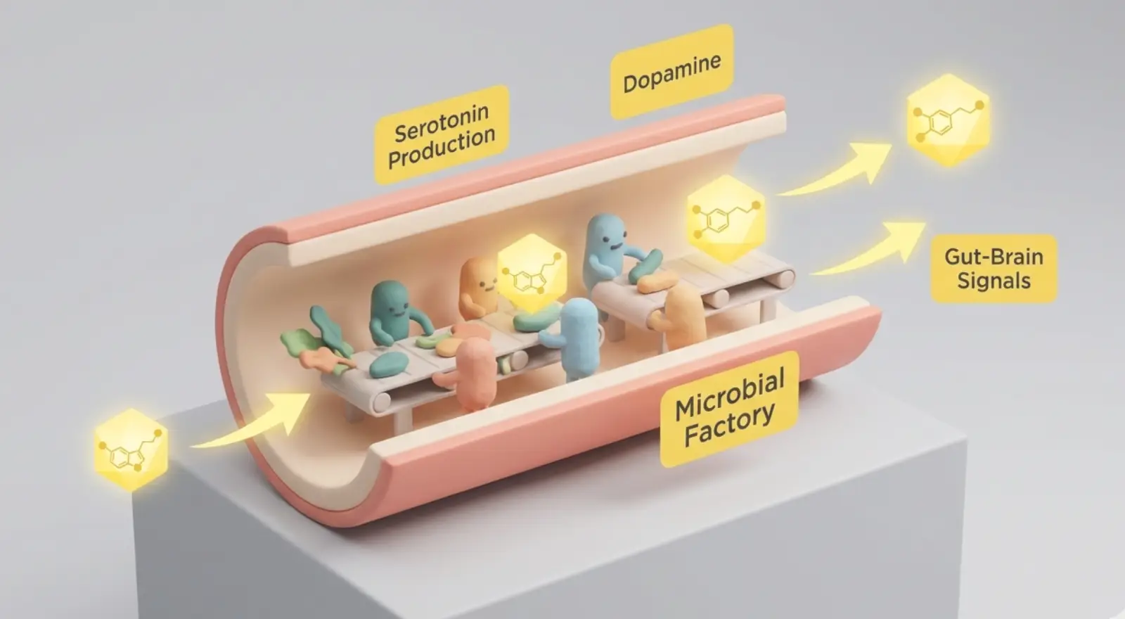
The gut-brain axis is increasingly recognized as a sophisticated system through which gut-derived signals—neural, microbial, and hormonal—influence brain function and memory. Multiple converging lines of research highlight the interplay between vagal signaling, microbiota, metabolic hormones, and their combined impact on hippocampal-dependent cognition.
Vagus Nerve Communicates Nutrition to Hippocampus
The vagus nerve is traditionally associated with calming the body, but its ascending fibers serve an excitatory purpose, relaying critical signals from the gut to the brain. Mechanoreceptors in the stomach monitor distension while chemoreceptors in the intestines sense nutrients, resulting in a complex “song” of signals rather than a single unified input. These diverse neural cues, transmitted via the vagus through the nodose ganglia to the brainstem, then reach the medial septum and finally the hippocampus, with acetylcholine as the primary neuromodulatory “currency” in this axis.
Vagus Nerve Lesioning Impairs Hippocampal-Dependent Memory via Gut-brain Pathway Disruption
Experimental lesioning of the gastrointestinal-innervating vagus nerve in animals leads to impairments in hippocampal-dependent memory tasks. Furthermore, vagal nerve stimulation (VNS) linked to natural feeding cues can rescue diet-induced memory impairments. Ongoing research explores whether VNS may ameliorate early Alzheimer’s pathology. The integrity of the vagal pathway is thus essential for optimal memory function.
Taiwanese Data Links 1980s Truncal Vagotomy For Ulcers to Increased Dementia Risk
Supporting data from Taiwan reveals that patients who underwent truncal vagotomy for ulcers in the 1980s-1990s show a higher probability of developing dementia later in life. This correlation implicates disrupted vagal signaling as a long-term risk factor for neurodegeneration. Ongoing registry analyses aim to further clarify these risks in different populations.
Acetylcholine in Hippocampus During Eating Encodes Context and Promotes Memory
Acetylcholine release in the hippocampus is dynamically modulated by eating. Using fiber photometry, studies reveal a “staircase” pattern: acetylcholine ramps up during eating, dips during pauses, and ends at a higher baseline post-meal. This postprandial acetylcholine elevation is thought to encode contextual memory of the meal, enabling animals to recall where and what they ate for future foraging success.
Taste and Post-Oral Nutrient Sensing Drive Vagal-Acetylcholine-Memory Signal
Post-ingestive nutrient sensing—not taste alone—is necessary for this gut-brain-memory pathway. Non-nutritive sweeteners like stevia, despite providing sweet taste, do not trigger hippocampal acetylcholine release in animal models. Instead, a combination of taste and nutrient detection after ingestion is essential to drive vagal signals that ultimately release acetylcholine in the hippocampus, indicating an integrated detection system that links sensory and metabolic cues for memory encoding.
Hormones Independently Signal Metabolic Status To Memory Systems
Beyond neural routes, the hippocampus hosts receptors for key metabolic hormones such as [restricted term], ghrelin, and leptin. These hormones deliver humoral signals about the body's energy status, providing a parallel input to the hippocampus alongside neural vagal signals. The hippocampus integrates these multisensory cues—neural and hormonal—to comprehensively monitor nutritional state and adapt memory processes accordingly.
Bacterial Populations Correlate Wit ...
Here’s what you’ll find in our full summary
Gut-brain Axis: Vagal, Microbial, Hormonal Connections to Memory
Additional Materials
Clarifications
- The vagus nerve is a major nerve connecting the brain to many organs, including the gut, and carries sensory and motor signals. The nodose ganglia are clusters of nerve cell bodies on the vagus nerve that relay sensory information from the gut to the brainstem. The medial septum is a brain region involved in memory and learning that receives signals indirectly from the vagus nerve via the brainstem. This pathway helps transmit gut-derived information to the hippocampus, influencing cognitive functions.
- Mechanoreceptors are sensory cells that detect physical changes like stretching or pressure in the gut walls. Chemoreceptors sense chemical substances such as nutrients and acids within the digestive tract. Both receptor types send signals to the brain to inform it about the gut’s current state. This information helps regulate digestion and influences brain functions like memory.
- Acetylcholine is a chemical messenger that modulates how neurons communicate, influencing learning and memory. Calling it a "neuromodulatory currency" means it acts like a key signal that adjusts brain activity in response to gut inputs. It enhances the hippocampus's ability to encode and store memories by changing neural circuit dynamics. This role is crucial for linking bodily states, like eating, to cognitive processes.
- Vagal nerve stimulation (VNS) involves delivering mild electrical pulses to the vagus nerve via an implanted device or external electrodes. When linked to feeding cues, VNS is timed to activate during or after eating, mimicking natural gut signals to the brain. This targeted stimulation enhances communication along the gut-brain axis, supporting memory processes affected by diet. Clinically, VNS is explored for treating neurological disorders by modulating brain activity through this pathway.
- Truncal vagotomy is a surgical procedure that involves cutting the main trunk of the vagus nerve to reduce acid secretion in the stomach and treat ulcers. This disrupts nerve signals between the gut and brain, affecting digestive functions and potentially other physiological processes. Because the vagus nerve also influences brain regions involved in memory, its severing may increase long-term risks like dementia. The procedure was more common before modern ulcer medications became available.
- Fiber photometry is a technique that uses light to measure activity of specific molecules like neurotransmitters in living brain tissue. It involves genetically encoding fluorescent sensors that change brightness when acetylcholine binds. A fiber optic cable implanted in the brain delivers excitation light and collects emitted fluorescence, allowing real-time monitoring. This method enables researchers to track neurotransmitter dynamics during behaviors such as eating.
- Taste sensing occurs in the mouth through taste buds detecting flavors like sweet, sour, or bitter. Post-ingestive nutrient sensing happens after food enters the gut, where specialized receptors detect actual nutrients such as sugars, fats, and amino acids. This gut sensing triggers hormonal and neural signals that inform the brain about the food’s nutritional value. Together, these systems help the body distinguish between mere flavor and real energy content.
- [restricted term] in the hippocampus supports memory by enhancing synaptic plasticity and energy metabolism. Ghrelin, known as the "hunger hormone," promotes memory formation by stimulating hippocampal neurons and neurogenesis. Leptin signals energy sufficiency and can improve learning and memory by modulating hippocampal synaptic activity. These hormones help the brain adjust cognitive functions based on the body's nutritional and energy status.
- Humoral signals are chemical messengers, like hormones, that travel through the bloodstream to reach target organs. Neural signals are electrical impulses transmitted directly between neurons via synapses. Unlike neural signals, humoral signals act more slowly and affect multiple tissues simultaneously. This allows the body to coordinate long-term physiological responses beyond immediate neural communication.
- Parabacteroides and Lacticoccus are types of gut bacteria that influence brain function by producing metabolites affecting neural pathways. Parabacteroides may release substances that impair memory-related brain regions, while Lacti ...
Counterarguments
- While animal studies provide compelling evidence for gut-brain axis mechanisms, direct causal links in humans remain less well established, and much of the evidence is correlational.
- The association between truncal vagotomy and increased dementia risk in Taiwanese patients may be confounded by other factors such as comorbidities, medications, or socioeconomic status, rather than vagal disruption alone.
- The effects of vagal nerve stimulation (VNS) on memory and Alzheimer’s pathology are still under investigation, and clinical efficacy in humans has not been conclusively demonstrated.
- The role of specific gut bacteria in memory function is complex and context-dependent; findings in animal models may not directly translate to humans due to differences in microbiome composition and environmental factors.
- The classification of bacteria as “beneficial” or “harmful” is highly context-specific and may oversim ...
Get access to the context and additional materials
Food Reward and Reinforcement: Plasticity, Preference, Dopamine System
Scott Kanoski discusses how food reward mechanisms in the brain are dynamic, deeply state-dependent, and shaped by physiological, psychological, and social influences—highlighting both specific neural circuits such as the mesolimbic [restricted term] pathway and the plasticity of food preferences.
Mesolimbic [restricted term] Pathway Responds To Food Reward Based On State
The mesolimbic [restricted term] system serves as a neural currency for multiple rewards, including both palatable foods and substances of abuse. Kanoski notes that there is considerable overlap in the brain between substance abuse and food consumption, but this does not mean food addiction is identical to drug addiction.
Palatable Foods Activate [restricted term] Release, but Bland Foods Can Do So When an Animal Is Food-Restricted or Nutrient-Deprived
Typically, palatable foods like sweets or high-fat items activate [restricted term] release. However, in food-restricted or nutrient-deprived states, even bland foods trigger pronounced [restricted term] signaling, demonstrating that the system is modulated by physiological need and relevance.
Mesolimbic [restricted term]: A Neural Currency For Rewards, State and Relevance Dependent
Thus, the mesolimbic [restricted term] system flexibly tracks the reward value of foods based on the animal’s current state, emphasizing that food rewards are not fixed but highly context-dependent.
Food Preferences: Plastic and Rewirable Through Choices
The food reward system is malleable: with changing eating patterns or experience, preferences and neural responses shift.
Sugar Reduction Alters Sweetness Perception Through Reward System Plasticity
Research shows that reducing sugar intake can change how sweetness is perceived, reflecting reward system plasticity. The effect is underpinned by changes in [restricted term] signaling that calibrate the perceived intensity or desirability of sweet foods as experience changes.
[restricted term] Release to Sucrose Eliminated After Learning Sucrose Predicts Illness, Showing Food Reward Circuitry Malleability
A dramatic example of plasticity in the food reward system is conditioned taste aversion. If an animal learns that sucrose consumption predicts malaise—such as after pairing sucrose with an illness-inducing compound—[restricted term] release in response to sucrose consumption disappears. This demonstrates that learned associations can profoundly rewire the neural response to foods once considered rewarding.
The Distinction Between Hunger Suppression and Craving Reduction
Appetite suppression and reduction in food cravings involve different neural mechanisms, and interventions may have distinct effects depending on which system they target.
Appetite Suppression by Glp-1 Drugs Involves Vagal and Brainstem Mechanisms, While Reducing Cravings May Require Engaging Higher-Order Brain Regions Like the Prefrontal Cortex and Hippocampus, Potentially Limited by Blood-Brain Barrier Penetration
Kanoski discusses drugs like GLP-1 agonists that reduce appetite. These appear to work primarily through increased meal satiation via the gut-vagus-brainstem pathway. Whether these drugs affect higher-order brain regions such as the prefrontal cortex or hippocampus—areas involved in cognition, memory, and perhaps controlling food-related thoughts or “food noise”—remains unclear due to questions about blood-brain barrier penetration ...
Here’s what you’ll find in our full summary
Food Reward and Reinforcement: Plasticity, Preference, Dopamine System
Additional Materials
Clarifications
- The mesolimbic [restricted term] system is a brain circuit involving [restricted term]-producing neurons that connect the midbrain to the limbic system, especially the nucleus accumbens. It signals the motivational importance of stimuli, helping the brain assign value to rewards and drive goal-directed behavior. [restricted term] release in this pathway reinforces behaviors by creating a feeling of pleasure or reward. This system adapts based on experience, influencing learning and decision-making related to rewards.
- [restricted term] is a chemical messenger in the brain that signals pleasure and motivation. When released, it reinforces behaviors by making them feel rewarding, encouraging repetition. This system helps the brain learn which actions lead to positive outcomes. [restricted term] release varies with context, reflecting the importance or desirability of a stimulus.
- Palatable foods are those that are highly flavorful and enjoyable, often rich in sugar, fat, or salt, which naturally stimulate [restricted term] release. Bland foods lack strong flavors or rewarding qualities and typically do not trigger significant [restricted term] responses under normal conditions. However, when an organism is hungry or nutrient-deprived, even bland foods become more valuable and can activate [restricted term] pathways. This shift reflects the brain’s adaptive mechanism to prioritize energy acquisition based on physiological need.
- "Food-restricted" means an organism is deliberately given less food than usual, leading to reduced energy intake. "Nutrient-deprived" refers to lacking specific essential nutrients like vitamins, minerals, or macronutrients despite possibly having some food. Both states trigger biological signals indicating energy or nutrient shortage, activating survival mechanisms. These signals alter brain chemistry, increasing the reward value of available foods to motivate eating.
- Neural plasticity refers to the brain's ability to change its structure and function in response to experience. In food preferences, this means the brain's reward circuits can adapt based on what foods are consumed and the outcomes associated with them. These changes alter how rewarding or desirable certain foods feel, influencing future choices. This adaptability helps organisms adjust their eating behavior to changing environments and internal states.
- Conditioned taste aversion occurs when an animal associates a specific food flavor with a negative experience, like illness, leading to avoidance of that food. This learning changes the brain’s reward response, so [restricted term] release triggered by that food decreases or stops. [restricted term] signaling normally signals pleasure or reward, so its reduction reflects the food becoming undesirable. This process shows how experiences can rewire neural circuits to alter food preferences.
- Appetite suppression primarily involves brainstem and vagal pathways that signal fullness and reduce the physical drive to eat. Craving reduction engages higher brain regions like the prefrontal cortex and hippocampus, which regulate motivation, desire, and cognitive control over food-related thoughts. These higher regions influence decision-making and emotional responses to food cues, beyond mere hunger signals. Effective craving reduction often requires drugs or interventions that can cross the blood-brain barrier to reach these areas.
- GLP-1 agonists mimic the hormone glucagon-like peptide-1, which is released after eating to promote fullness. They activate receptors on vagal nerve fibers in the gut, sending signals to the brainstem to reduce hunger and slow stomach emptying. This gut-brain communication decreases meal size by enhancing feelings of satiation. The pathway operates largely outside conscious control, influencing appetite through physiological feedback.
- The blood-brain barrier (BBB) is a protective layer of tightly joined cells that prevents many substances in the bloodstream from entering the brain. It selectively allows essential nutrients to ...
Counterarguments
- While the mesolimbic [restricted term] system is implicated in both food and drug rewards, equating their mechanisms may oversimplify the unique complexities and consequences of substance use disorders compared to eating behaviors.
- The assertion that food reward is highly context-dependent may understate the influence of genetic predispositions or long-term habitual patterns that can persist despite changes in physiological state.
- Evidence for the plasticity of food preferences is robust, but some individuals exhibit persistent preferences or aversions that are resistant to change, suggesting limits to neural and behavioral plasticity.
- The impact of reducing sugar intake on sweetness perception may vary widely among individuals, and not all people experience significant recalibration of reward responses.
- Conditioned taste aversion is a powerful model in animals, but its translation to human eating behavior is less direct, as humans often continue to consume foods associated with negative experiences.
- The distinction between appetite suppression and craving reduction is supported by some evidence, but in practice, these processes often overlap and may not be entirely separable in real-world interventions.
- The effectiveness of GLP-1 agonists in reducing cravings for u ...
Get access to the context and additional materials


Create Summaries for anything on the web
Download the Shortform Chrome extension for your browser